• 01 49 80 97 97
  • Contact us
  • Boutique NIDEK

Efficient After Sales Service

At your disposal whatever the situation

Made in France

Our consultation units

Our experts at your disposal

60 experts over the territory

  • Lasers
        • Lasers YAG and SLT
          • YC-200 S Plus (YAG/SLT)
          • YC-200 (YAG)
        • Photocoagulator lasers
          • YLC-500 / YLC 500 VIXI
          • GYC-500 / GYC 500 VIXI
  • Retina diagnosis
        • Slit lamps
          • SL-9900/SL-9900 Zoom
          • SL-9800
          • SL-250
        • Multimodal platforms
          • Mirante (SLO/OCT)
          • Mirante SLO (SLO)
        • Fundus cameras
          • AFC-330
          • Retina Scan Duo 2
          • Mirante SLO (SLO)
          • Mirante (SLO/OCT)
        • Microperimeters
          • MP-3
          • MP-3S
        • Angiographs
          • Mirante (SLO/OCT)
          • Mirante SLO (SLO)
        • Software
          • NAVIS-EX Software
        • OCT
          • RS-1
          • Retina Scan Duo 2
          • Mirante (SLO/OCT)
        • Ultrasound scanners
          • US-4000/4000-P (Echo A+B)
          • US-500 / 500-P (Echo A)
  • Anterior segment
        • Slit lamps
          • SL-9900/SL-9900 Zoom
          • SL-9800
          • SL-250
        • Aberrometers - Topographs
          • OPD-Scan III
        • Optical biometers
          • AL-Scan
          • AL-Scan M
        • Tonometers
          • TONOREF III
          • NT-1e / NT-1 / NT-1p
        • Specular microscope
          • CEM-530
        • Goniolenses
          • GS-1
  • Refraction
        • Pupillometers
          • PM-700
        • Afficheurs de tests
          • CP-9
          • SC-1600 Pola/SC-1600
        • Itinerant optician case
          • Itinerant optician case
        • Lensometers
          • LM 1800 P – PD
          • LM7 – 7P
        • Auto refractometers
          • AR-1/ARK-1
          • AR-1a/ARK-1a
          • AR-1s/ARK-1s
          • AR-F/ARK-F
          • HandyRef/HandyRef-K
          • TONOREF III
        • Refractors
          • RT-3100
          • TS-310
          • RT-6100
          • TS-610
          • Logiciel RT-Connect
        • Système de réfraction assisté
          • FARS (Fully Assisted Refraction System)
  • Consultation units
        • "Excellence" units
          • Synetic
          • OT-6400
        • "Performance" units
          • Affinity
          • OT-4200
        • "Compact" units
          • ONE-i
          • Lypop
          • COS-6100
        • Lifting tables and examination swivel chair
          • Consultation armchairs and stools
          • Tables à élévation Up
  • Optical workshop
        • Automatic edges
          • ME-1500
          • LEXCE Trend 8D
          • LEXCE Trend D
          • LEXCE Trend DBT
          • SE9090 Supra
        • Lensometers
          • LM 1800 P – PD
          • LM7 – 7P
        • Centering-blocking devices
          • ICE-1500
          • ICE- 1
        • Shape plotters
          • LT-980
          • LETR-3 Traceur LEXCE Trend
        • Green products
          • LFU-220
          • LED-200 Plus
        • Colonne de centrage : Invision Fit AI
  • NIDEK
        • Our services
          • Design office
          • Installation & Implementation
          • Trainings
          • ASS and repairing
          • Environement
          • Showroom NIDEK
        • Our solutions
          • Optics
          • Ophthalmology
          • Orthoptics
          • Health care establishment
        • About us
          • Expertise and know-how
          • Certifications
          • NIDEK in France
          • NIDEK worldwide
          • Recruitment
        • News
          • Blog
          • Évènements
  • Our shop
  • fr_FR
  • en_GB

Homepage > Ultrasound scanners

Ultrasound scanners

Diagnostic rétine et segment antérieur
Our products
Benefits
Getting equipped
AN ADAPTED MEASURE

Additional imaging

The ultrasound scanning is a biometry and imaging examination using an ultrasound sound beam. On one hand, it makes possible measuring the different eye-structure lengths (axial length, cornea or crystalline lens thickness) and on the other hand rebuilding a 2D view (section) of the eye, particularly the fundus (retina). It can also be used to observe the tissues around the eye and the orbit.

Il est principalement utilisé lorsque les milieux internes de l’œil ne sont plus assez transparents (cataracte dense, hémorragies, inflammations…) et empêchent le passage correct de la lumière (utilisée par les biomètres optiques ou la lampe à fente).

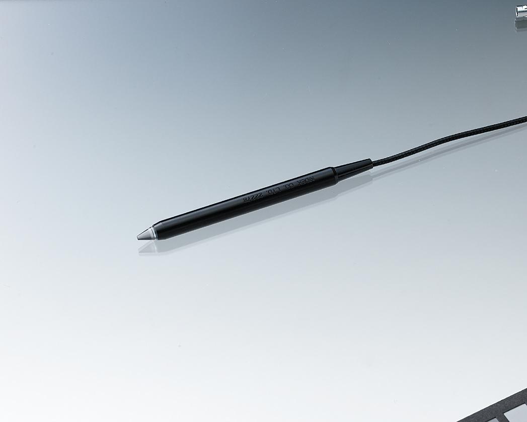
Depending on the examination mode, there is a contact with cornea (echo A) of the eyelid (echo B). The doctor has to be careful to correctly perform the examination. Using a different probe, it is also possible to measure the cornea thickness, as it a precious value to follow-up glaucomas or detect some pathologies. In this case, the probe is applied on the cornea.

NIDEK PRODUCTS

Nos solutions d'échographes

Loading...

US-4000/4000-P (Echo A+B)

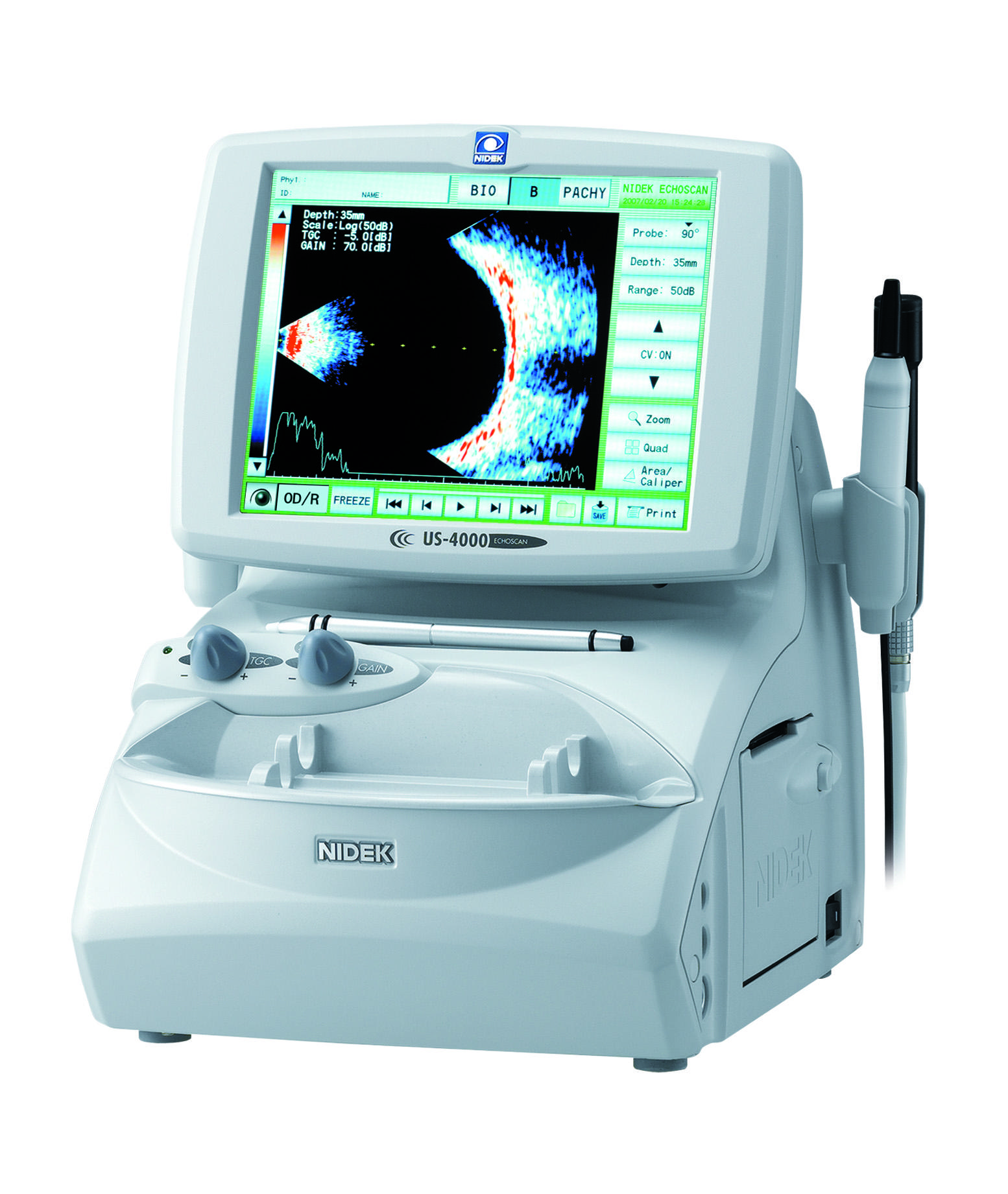
The ultrasound scanner US-4000 proposes eye biometric analyses, with the measure of the axial length, thanks to the ultrasound technology in mode echo-A, and 2D sections of the eye in mode echo-B. The option US-4000P is, in addition, equipped with a probe dedicated to measuring the thickness of the cornea, or pachymetry.

FIND MORE

US-500 / 500-P (Echo A)

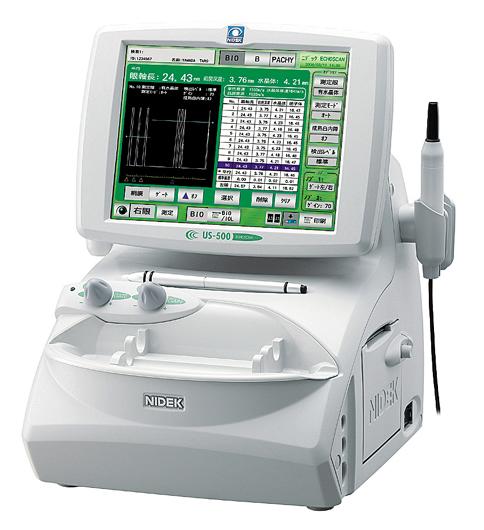
The ultrasound scanner US-500 proposes eye biometric analyses, and particularly the axial length, thanks to the ultrasound technology in mode echo-A. The option US-4000P is, in addition, equipped with a probe dedicated to measuring the thickness of the cornea, the pachymetry.

FIND MORE
NIDEK EXPERTISE

Techniques used

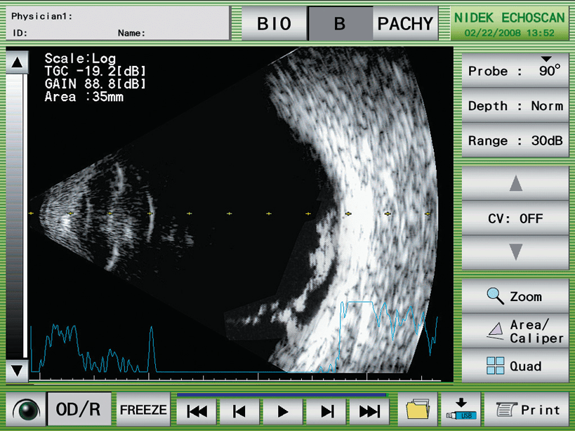
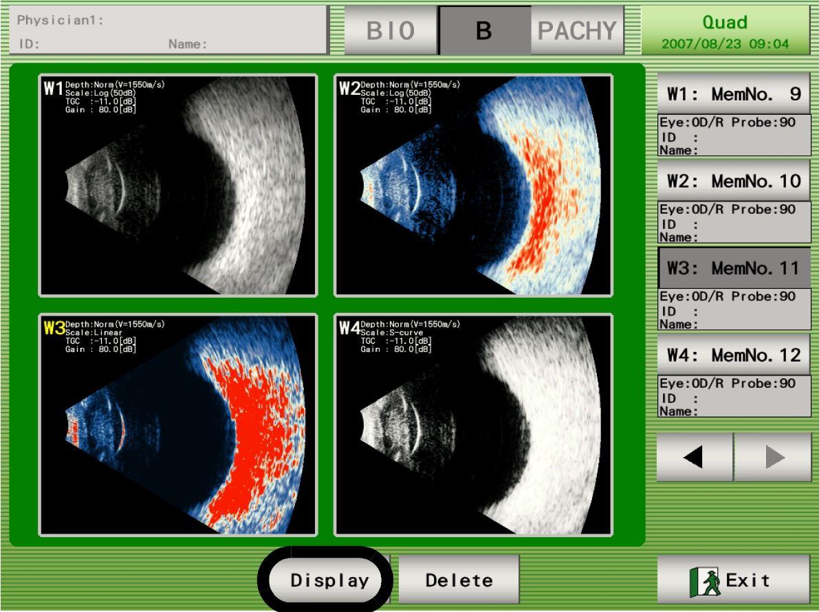
Ultrasound scanning is an examination using a high-frequency ultrasound sound beam the human ears cannot hear. Thanks to the reflexions of the beam, resulting from structure-composition differences produced when the beam goes through the eye, the ultrasound scanner collects echos to calculate anatomical lengths with a high precision or to rebuild a 2D image (section) using two-dimensional scanning.

As the sound propagation velocity in the different media of eye are known, the lengths are measured depending on the echo RTT.

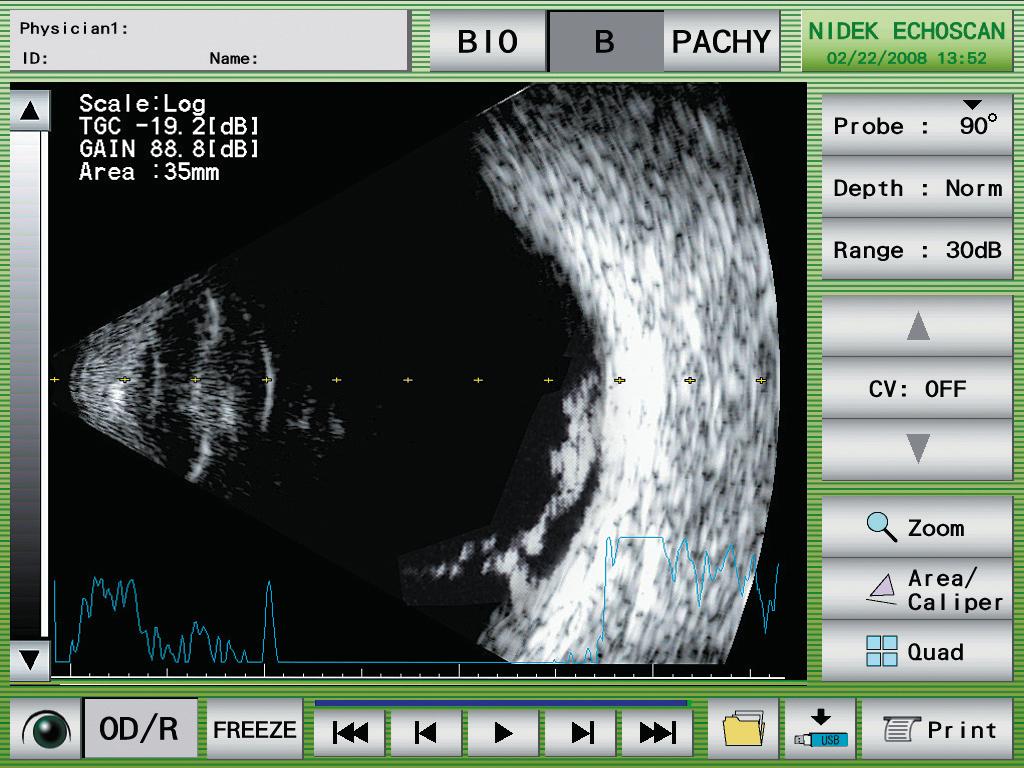
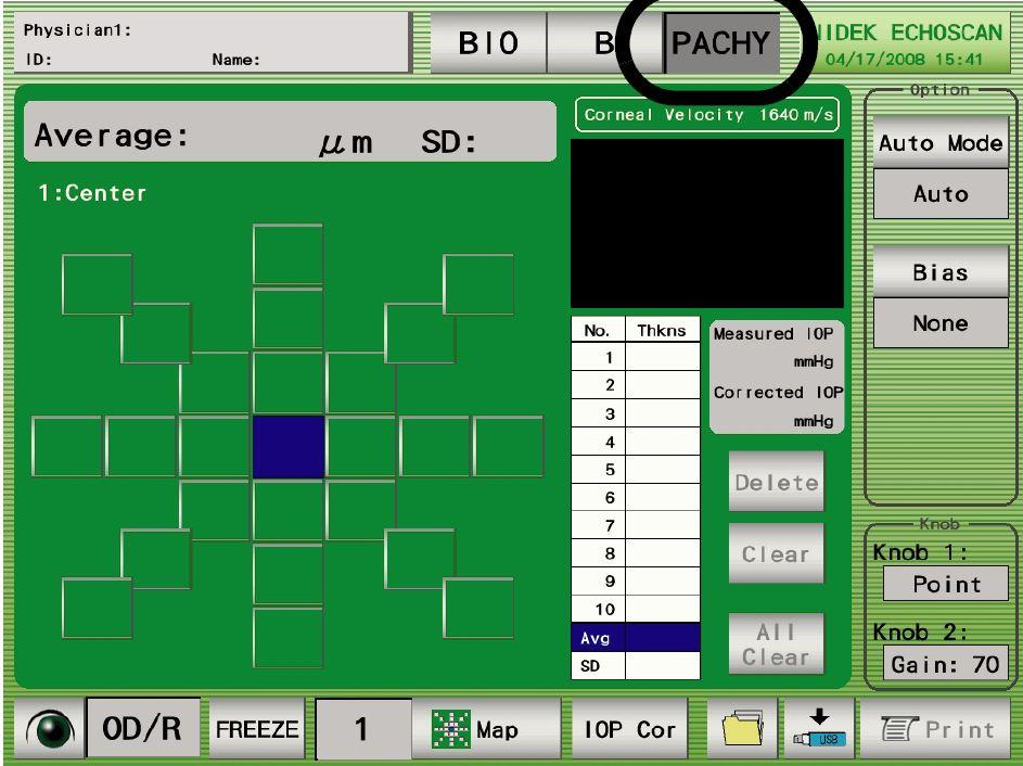
If the sound beam scans a 2D surface, echos are assembled to rebuild a 2D image, i.e. a section of the probed structure. As it is not limited by haemorrhages and opacities, the ultrasound scanner is the perfect choice when optical instruments using a light beam are ineffective. It is particularly recommended in case of eye tumours or inflammations. Thanks to a special probe, measuring the thickness of the cornea, or pachymetry, is also possible. This examination means setting the probe on the patient’s cornea, but the value is useful in case a glaucoma is suspected or when scheduling a surgery.

MODE A VS MODE B

The different types of ultrasound scanning

Depending on the probe and the scanning mode of the eye, there are two ultrasound-scanning techniques. The purposes and the medical applications are therefore different.

Mode A

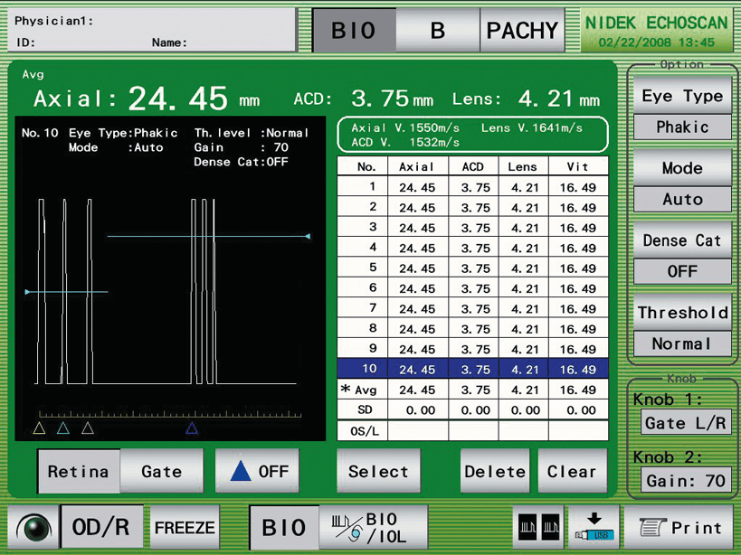
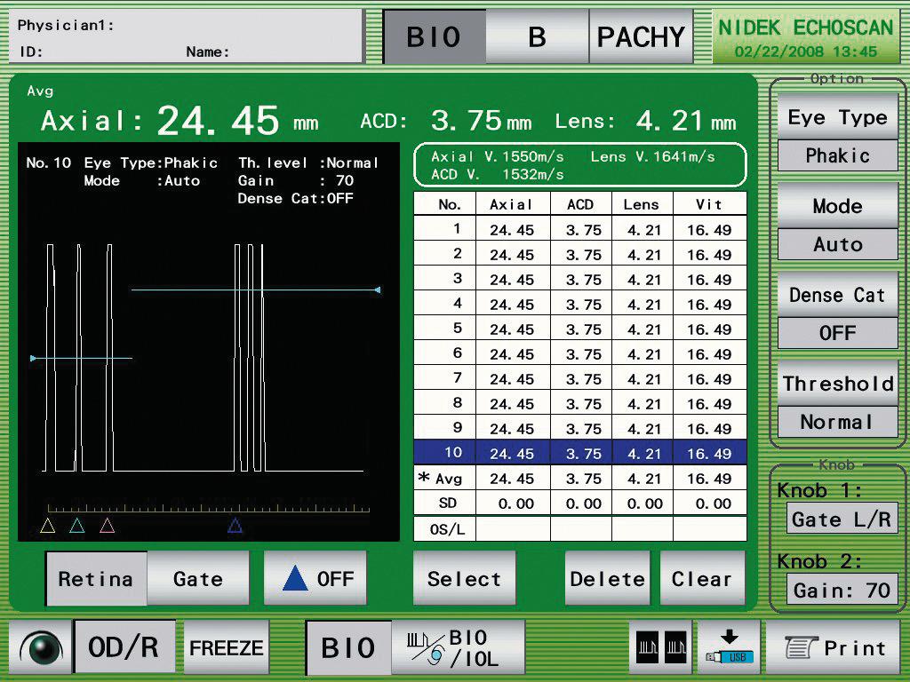
In mode A, only one sound beam is sent through the eye and only its echos are detected. It is then possible to measure the anatomic distances of eye structures (eye total length, crystalline lens or cornea thickness, anterior chamber length, etc.). Mode-A ultrasound scanning mainly applies to the calculation of the intraocular implant, artificial implant replacing the crystalline lens after a cataract surgery, when optical biometers are limited.

Mode B

In mode B, or two-dimensional ultrasound scanning, the sound beam scans a larger part of the eye and the different echo signals are used to rebuild a 2D image. Mode-B ultrasound scanning is often used when the fundus cannot be viewed using a slit lamp (opacity of the eye internal media), in case of eyeball traumatism, uveitis, inflammation, eye tumour or any pathology creating a haemorrhage hiding fundus.

Benefits

Using a sound signal the human being cannot hear, the eye ultrasound scanning examination is a non (or low) invasive procedure providing a biometry and an eye analysis for difficult cases when usual instruments are ineffective.
  • Implant calculation

Eye biometry with calculation function of the implant, for cataract surgeries.

  • Additional imaging

In mode B, visualisation of the fundus in case of haemorrhage or opaque media.

  • Pachymetry

Option designer to assist glaucoma diagnosis, surgery or the detection of some pathologies.

Vous souhaitez vous équiper d'un échographe ?

You have a project? You want a quotation? You have questions about our products? Feel free to ask your technical sales representative.

Contact us

The Art of Eye Care

Reliability and
safety

NIDEK develops its top-of-the-range products to improve visual health through an approach based on strict criteria: safety, reliability, durability, continuous quality controls and certifications.

Technologies and
innovations

NIDEK meets technical challenges by keeping constantly informed of the innovations of eye imaging systems, using the expertise of professionals and the progresses of research.

Services and
guarantees

NIDEK commits itself to providing services to its customers, from the installation of an activity to the authorised training of teams, and to offering long-time measurable guarantees.

FIND MORE
Le magazine

L'actualité NIDEK

Notre actualité

Les OCT Nidek référencés par l’URPS

Les OCT Nidek référencés par l’URPS : Une opportunité pour les professionnels de santé visuelle

Lire la suite »

🎉 NIDEK à la SFO 2025 : notre gamme médicale à l’honneur !

🎉 NIDEK à la SFO 2025 : notre gamme médicale à l’honneur ! Du 10

Lire la suite »

NIDEK accompagne la formation des assistants médicaux en ophtalmologie avec Keyce Santé

Chez NIDEK, nous sommes convaincus que l’innovation ne se limite pas aux équipements : elle

Lire la suite »

Retour sur la visite de M. Ozawa, président de NIDEK, en France

Le 11 février, NIDEK SA France a eu l’honneur d’accueillir M. Ozawa, président de NIDEK,

Lire la suite »
Découvrir le blog
Nos événements

🎉 NIDEK à la SFO 2025 : notre gamme médicale à l’honneur !

🎉 NIDEK à la SFO 2025 : notre gamme médicale à l’honneur ! Du 10

Lire la suite »

Retour sur la visite de M. Ozawa, président de NIDEK, en France

Le 11 février, NIDEK SA France a eu l’honneur d’accueillir M. Ozawa, président de NIDEK,

Lire la suite »

Nidek et MEDADOM renouvellent leur partenariat pour leur dispositif innovant de téléophtalmologie

Communiqué de presse Nidek et MEDADOM renouvellent leur partenariat pour leur dispositif innovant de téléophtalmologie

Lire la suite »

NIDEK SA à la Journée Nationale de Téléophtalmologie 2024

Nous sommes ravis d’annoncer la participation de NIDEK SA à la Journée Nationale de Téléophtalmologie

Lire la suite »
Tous nos événements

Contact NIDEK

Contact us

Spécialiste des équipements et technologies au service de la santé visuelle, NIDEK SA accompagne et équipe les ophtalmologistes, orthoptistes, professionnels de l’optique et les établissements de santé.

Facebook Linkedin Youtube

KEY PRODUCTS

  • Mirante
  • TS 610
  • RT-Connect
  • Synetic

Services

  • Design office
  • Installation & Implementation
  • Trainings
  • ASS and repairing

Solutions

  • Optics
  • Ophthalmology
  • Orthoptics
  • Health care establishment

About us

  • Expertise and know-how
  • Our key dates
  • Certifications
  • Nidek in France and worldwide
  • Recruitment

© 2021 • NIDEK SA – All rights reserved

  • General Terms and Conditions
  • Protection des données personnelles
  • Legal notices
Gérer le consentement aux cookies
Nous utilisons des cookies pour optimiser notre site web et notre service.
Fonctionnel Always active
Le stockage ou l’accès technique est strictement nécessaire dans la finalité d’intérêt légitime de permettre l’utilisation d’un service spécifique explicitement demandé par l’abonné ou l’utilisateur, ou dans le seul but d’effectuer la transmission d’une communication sur un réseau de communications électroniques.
Préférences
Le stockage ou l’accès technique est nécessaire dans la finalité d’intérêt légitime de stocker des préférences qui ne sont pas demandées par l’abonné ou l’utilisateur.
Statistiques
Le stockage ou l’accès technique qui est utilisé exclusivement à des fins statistiques. Le stockage ou l’accès technique qui est utilisé exclusivement dans des finalités statistiques anonymes. En l’absence d’une assignation à comparaître, d’une conformité volontaire de la part de votre fournisseur d’accès à internet ou d’enregistrements supplémentaires provenant d’une tierce partie, les informations stockées ou extraites à cette seule fin ne peuvent généralement pas être utilisées pour vous identifier.
Marketing
Le stockage ou l’accès technique est nécessaire pour créer des profils d’utilisateurs afin d’envoyer des publicités, ou pour suivre l’utilisateur sur un site web ou sur plusieurs sites web ayant des finalités marketing similaires.
Manage options Manage services Manage {vendor_count} vendors Read more about these purposes
Préférences
{title} {title} {title}

Télécharger votre brochure

Télécharger votre brochure

Télécharger votre brochure

Télécharger votre brochure

Télécharger votre brochure

Télécharger votre brochure

Télécharger votre brochure

Télécharger votre brochure

Télécharger votre brochure

Télécharger votre brochure

Télécharger votre brochure

Télécharger votre brochure

Télécharger votre brochure

Télécharger votre brochure

Télécharger votre brochure

Télécharger votre brochure

Télécharger votre brochure

Télécharger votre brochure

Télécharger votre brochure

Télécharger votre brochure

Télécharger votre brochure

Télécharger votre brochure

Télécharger votre brochure

Télécharger votre brochure

Ce site est réservé aux professionnels de santé

Sortir
Je suis professionnel de santé