• 01 49 80 97 97
  • Contact us
  • Boutique NIDEK

Efficient After Sales Service

At your disposal whatever the situation

Made in France

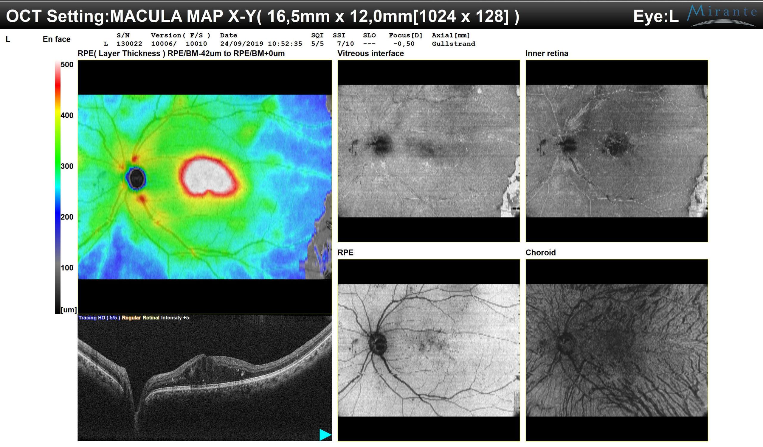
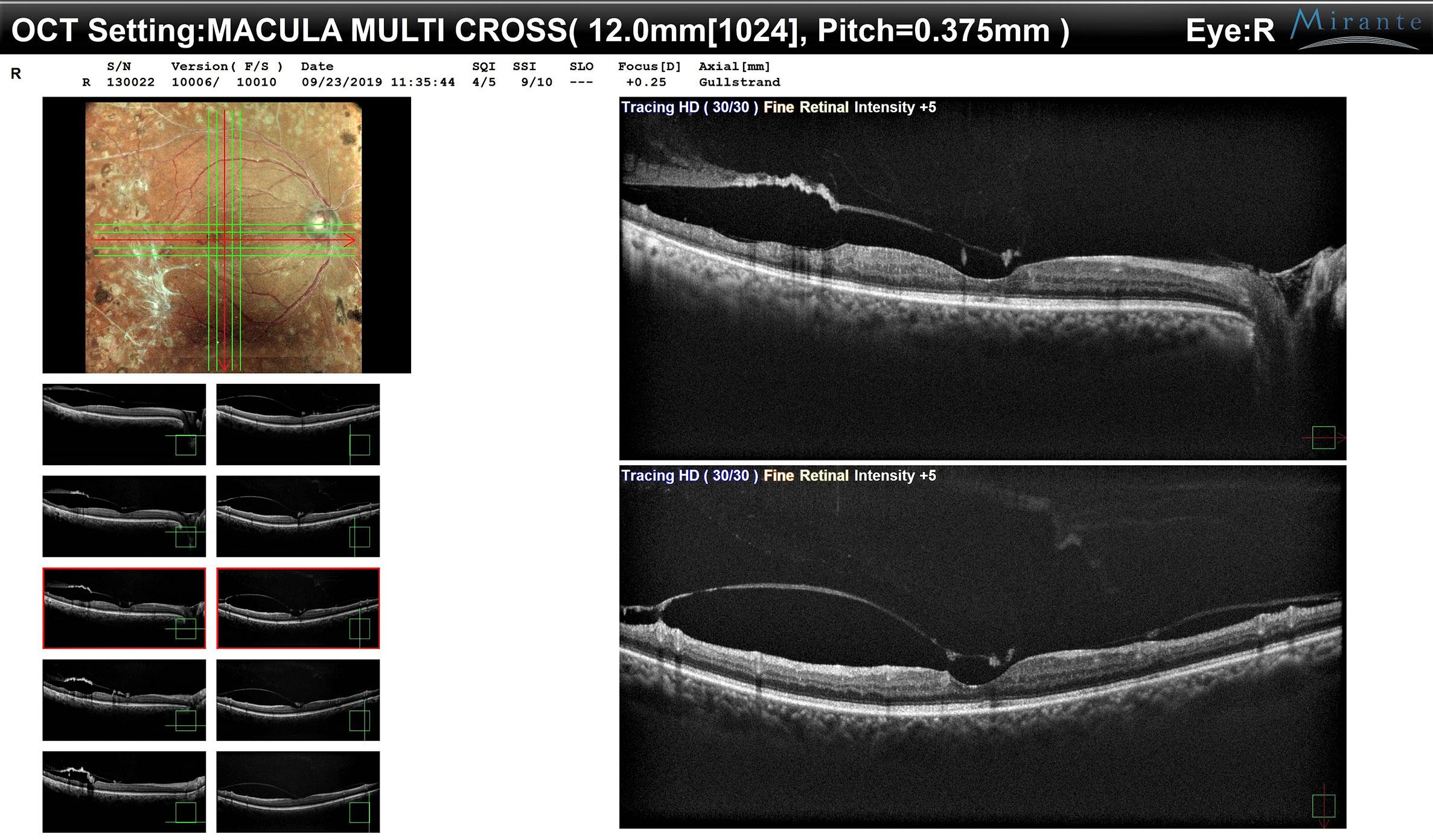
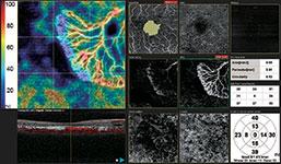
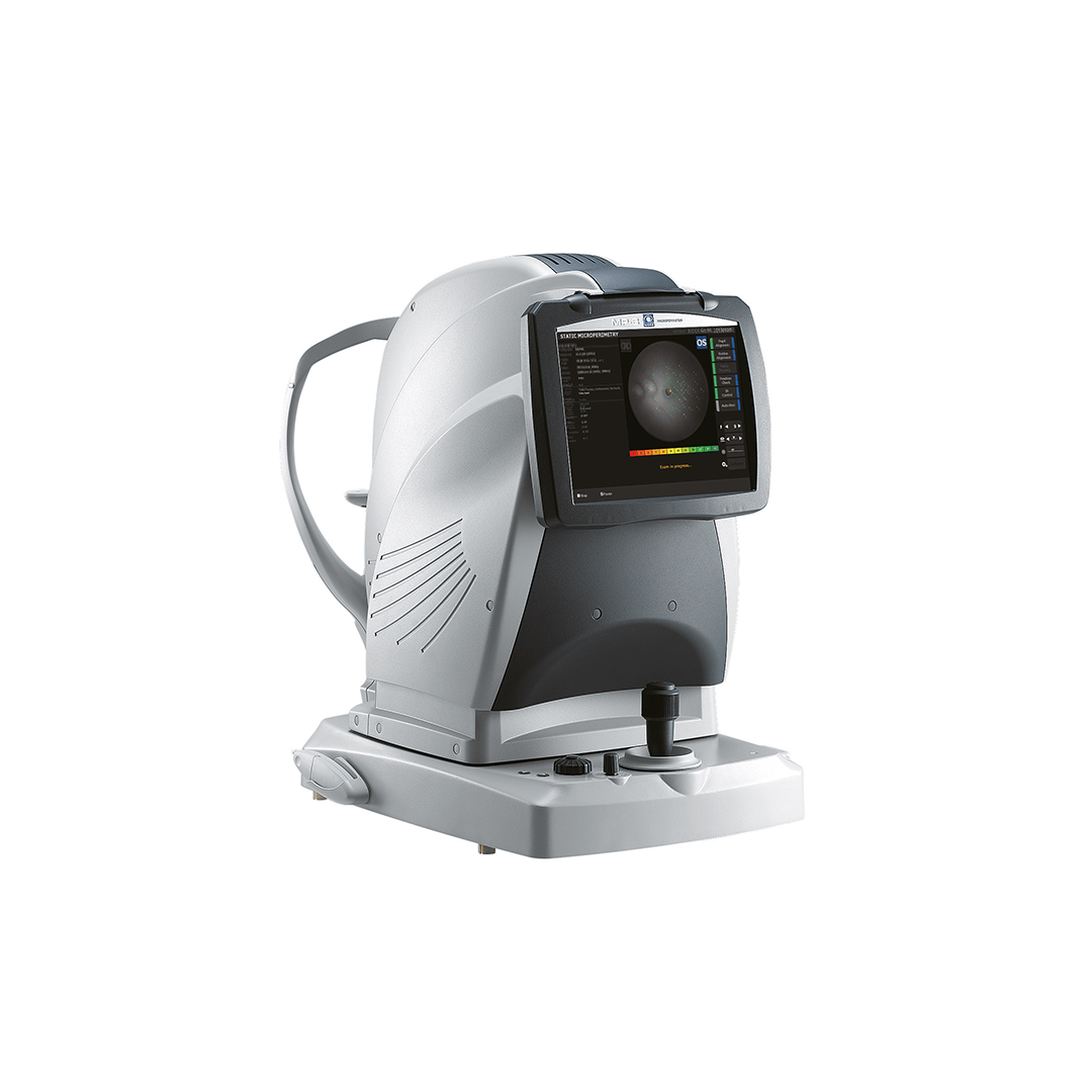
Our consultation units

Our experts at your disposal

60 experts over the territory

  • Lasers
        • Lasers YAG and SLT
          • YC-200 S Plus (YAG/SLT)
          • YC-200 (YAG)
        • Photocoagulator lasers
          • YLC-500 / YLC 500 VIXI
          • GYC-500 / GYC 500 VIXI
  • Retina diagnosis
        • Slit lamps
          • SL-9900/SL-9900 Zoom
          • SL-9800
          • SL-250
        • Multimodal platforms
          • Mirante (SLO/OCT)
          • Mirante SLO (SLO)
        • Fundus cameras
          • AFC-330
          • Retina Scan Duo 2
          • Mirante SLO (SLO)
          • Mirante (SLO/OCT)
        • Microperimeters
          • MP-3
          • MP-3S
        • Angiographs
          • Mirante (SLO/OCT)
          • Mirante SLO (SLO)
        • Software
          • NAVIS-EX Software
        • OCT
          • RS-1
          • Retina Scan Duo 2
          • Mirante (SLO/OCT)
        • Ultrasound scanners
          • US-4000/4000-P (Echo A+B)
          • US-500 / 500-P (Echo A)
  • Anterior segment
        • Slit lamps
          • SL-9900/SL-9900 Zoom
          • SL-9800
          • SL-250
        • Aberrometers - Topographs
          • OPD-Scan III
        • Optical biometers
          • AL-Scan
          • AL-Scan M
        • Tonometers
          • TONOREF III
          • NT-1e / NT-1 / NT-1p
        • Specular microscope
          • CEM-530
        • Goniolenses
          • GS-1
  • Refraction
        • Pupillometers
          • PM-700
        • Afficheurs de tests
          • CP-9
          • SC-1600 Pola/SC-1600
        • Itinerant optician case
          • Itinerant optician case
        • Lensometers
          • LM 1800 P – PD
          • LM7 – 7P
        • Auto refractometers
          • AR-1/ARK-1
          • AR-1a/ARK-1a
          • AR-1s/ARK-1s
          • AR-F/ARK-F
          • HandyRef/HandyRef-K
          • TONOREF III
        • Refractors
          • RT-3100
          • TS-310
          • RT-6100
          • TS-610
          • Logiciel RT-Connect
        • Système de réfraction assisté
          • FARS (Fully Assisted Refraction System)
  • Consultation units
        • "Excellence" units
          • Synetic
          • OT-6400
        • "Performance" units
          • Affinity
          • OT-4200
        • "Compact" units
          • ONE-i
          • Lypop
          • COS-6100
        • Lifting tables and examination swivel chair
          • Consultation armchairs and stools
          • Tables à élévation Up
  • Optical workshop
        • Automatic edges
          • ME-1500
          • LEXCE Trend 8D
          • LEXCE Trend D
          • LEXCE Trend DBT
          • SE9090 Supra
        • Lensometers
          • LM 1800 P – PD
          • LM7 – 7P
        • Centering-blocking devices
          • ICE-1500
          • ICE- 1
        • Shape plotters
          • LT-980
          • LETR-3 Traceur LEXCE Trend
        • Green products
          • LFU-220
          • LED-200 Plus
        • Colonne de centrage : Invision Fit AI
  • NIDEK
        • Our services
          • Design office
          • Installation & Implementation
          • Trainings
          • ASS and repairing
          • Environement
          • Showroom NIDEK
        • Our solutions
          • Optics
          • Ophthalmology
          • Orthoptics
          • Health care establishment
        • About us
          • Expertise and know-how
          • Certifications
          • NIDEK in France
          • NIDEK worldwide
          • Recruitment
        • News
          • Blog
          • Évènements
  • Our shop
  • fr_FR
  • en_GB
PageProduit_MiranteComplet_VisuelProduit_Proposition1

Homepage > Multimodal platforms > Mirante (SLO/OCT)

Mirante (SLO/OCT)

The Mirante is a large-field imaging multimodal platform designed to fundus imaging. It combines OCT and LSO imaging, thus grouping all routine imaging to detect and identify retinal-choroidal pathologies. By adding a lens, performing a section analysis of the anterior segment is possible.

  • More details
  • Vidéos du produit
Call me back
Download the brochure
  • Summary
  • Description
  • Accessories
  • Cleaning
  • Documentation
  • LSO confocal imaging
    • 4 laser sources and 4 dedicated sensors (blue 488 nm, green 532 nm, red 670 nm and IR 790 nm)
    • Standard 89° acquisition field
    • Ultra large 163° acquisition field
    • Image still resolution up to 4096×4096 pixels
    • Video resolution up to 1024×1024 pixels
    • FlexTrack algorithm to correct distortions
    • Taking into account cyclotorsion
  • OCT SD imaging at 85,000 A-scans/s
    • Acquisition field up to 16x5x12 mm
    • HD mode up to 120 sections
    • Real-time automatic repositioning of scans
    • Taking into account cyclotorsion
    • Follow-up upon acquisition
    • COMBO mode to delegate the tasks
    • OCT-Angiography
    • EDI mode for the choroid
    • Normative database over 9x9 mm for the macula and over 6x6 mm for the papilla zone
    • Extra lens for the OCT sections of the anterior segment
    • OCT B-Scan Denoising Software
  • Dioptrical compensation from -15D to + 15D
  • Internal red or blue fixation point and external white fixation points.
  • 100% customisable examination reports

A complete imaging tool of the retina

The Mirante embraces in a single product all the imaging modalities used daily to observe the fundus. They can also be obtained in ultra large field mode:

  • Colour photography
  • Deconstruction in a single Red/Green/Blue colour
  • IR photography
  • Auto fluorescence (FAF-Blue/FAF-Green)
  • Retro mode
  • FA and ICG dynamic and static angiographies
  • OCT
  • OCT-Angiography

It integrates a LSO (Laser Scanning Ophtalmoscope) that uses infrareds and provides high-contrast and high-resolution images. This systems also performs the tracking of the patient’s eye before and during the OCT mode acquisition, thus ensuring the correct positioning of OCT and OCT-Angiography scans by considering the natural cyclotorsion of human eye.

PageProduit_MiranteComplet_VisuelProduit_Proposition2
Image IR SLO
LSO IR Image
Image couleur
Image couleur
Tracking SLO en OCT
Tracking SLO en OCT
Image couleur – reconstruction à partir des trois canaux R/V/B
Image rouge – Choroïde
Red image – Choroid
Image verte – Réseau vasculaire (hémorragies, anévrismes…)
Image bleue – Tête du nerf optique et des fibres nerveuses
Autofluorescence Verte – Zone maculaire non masquée
Autofluorescence Bleue – Zone maculaire masquée mais tête du nerf optique plus visible
Ecran d’acquisition an angiographie simultanée FA et ICG avec IR en temps réel
PageProduit_MiranteComplet_Description_Visuel18_PhasesAngio
PageProduit_MiranteComplet_Description_Visuel19_CasRetro1_A
PageProduit_MiranteComplet_Description_Visuel19_CasRetro1_B
PageProduit_MiranteComplet_Description_Visuel20_CasRetro2_A
PageProduit_MiranteComplet_Description_Visuel20_CasRetro2_B
PageProduit_MiranteComplet_Description_Visuel23_PériphFA
PageProduit_MiranteComplet_Description_Visuel24_Panorama

LSO imaging of the fundus

The images of the fundus obtained through an LSO scanning and a normal 89° field, and a large field up to 163° are high-definition images, with a premium contrast.

The analysis of colour photos and red, green and blue original images, obtained directly and without a filter from each wavelength-dedicated sensor makes possible viewing the finest details of the fundus to easily detect the lesions arising in retinal-choroidal pathologies.

We use the auto fluorescence to analyse the pigmentary epythelium. There are two versions of auto fluorescence: the green wavelength one to correctly view the macula and the blue wavelength one, more convenient to observe the papilla. Therefore, adapting the reading to the studied pathology is possible.

Dynamic or static injection angiography acquisitions, using fluorescein and/or the ICG, are used to observe the vascularisation of the retina and the choroid, from the early filling phases to the late times. Thanks to the real-time infrared imaging, the alignment is performed at the beginning of the injection, thus reducing the loss of information. The automatic setting of the gain, independent for the fluorescein and the ICG, limits the saturation of the signal that sometimes hind the fundus. You can then confirm your diagnosis using the detection of vascular lesions, such as aneurysms, ischemia and leaks.

The retro mode is a technology specially attached to the Mirante. It is an infrared imaging of the deep layers of retina to highlight the structures of the pigmentary epythelium and of the choroid. It uses, derivatively, the LSO technology, and reinforces the detection of fine lesions on these difficult-to-access layers, particularly the lesions of drusen, the conventional imaging methods cannot sometimes see.

The ultra large field adapter is used to enlarge the analysis zone up to 163°, to access to the periphery. By tilting and rotating the upper part of the product, you can observe the fundus, up to the extreme periphery. The reconstruction of different pictures provides a view of the entire fundus, up to the ora serrata.

OCT and OCT-Angiography imaging

Designed to performed specialised consultations, the Mirante integrates different advanced OCT functions, such as the HD mode, the manual positioning of acquisition scans on the area of interest, the Select mode and Rescan, to perform an additional exam, or the following-up of patients, available as soon as the acquisition.

Thanks to the LSO Tracking, the Mirante can perform the following-up of examinations exactly in the same conditions, through the recognition of the patient’s retina. This ensures a reliable comparative analysis, including on a single section. Up to 50 examinations can be compared over time, to monitor the evolution of the disease and/or to control the efficiency of the cure. Use trend curves and add a manual event, such as the date of the first use of the therapy, for instance, to perform an efficient and tailor-made following-up

The Mirante also integrates an anterior segment adapter to make sections of the cornea and of the filtration angles.

The AngioScan makes possible the analysis of the retinal and choroidal vascularisation. The real time tracking of the retina ensures a correct positioning of the scans, to limit any motion artefacts. To make the analyses different, a picture is taken either of the macula (Macula Map), or of the optical nerve head (Disc Map). The segmentations are adapted to the captured zone, 7 different zones on the macula and 4 zones on the papilla, and the density and vascular perfusion values are given to perform an additional quantitative analysis.

Thanks to the combined OCT En-Face and OCT-Angiography display, whose depth can be adjusted, and to the B-scan section (from a drop-down menu), a direct matching can be made between the structure and the tissue vascularisation.

Examen de Follow-Up avec reconnaissance de la rétine et repositionnement du scan
Analyse Follow-Up comparative sur Macula Map, complexe des cellules ganglionnaires
Comparative follow-up analysis using Macula Map of the complex of ganglionic cells.
Cartographie d’épaisseur de la cornée
Mapping of cornea thickness
Mesure de l’angle irido-cornéen
Analyse OCT-A sur Macula – 7 segmentations avec valeurs de densité vasculaire et zone avasculaire centrale
Analyse OCT-A sur Papille – 4 segmentations avec valeurs de densité vasculaire
OCT-A analysis of the papilla – 4 segmentations with vascular density values.
Analyse comparative OCT En Face / OCT-A / B-scan sur Macula
Analyse comparative OCT En Face / OCT-A / B-scan sur Papille
Analyse OD/OG en photocouleur ultra grand champ
Analyse en photocouleur et décomposition R/G/B
Colour photo analysis and R/G/B deconstruction
Analyse comparative entre modalités
Rapport d’examen d’imagerie mutlimodale
Epaisseur de rétine sur 16,5 x 12 mm avec BDN sur 9x9mm
Analyse du cube 3D ligne par ligne (Macula)
Line-by-line 3D cube analysis (macula)
Cartographie RNFL avec BDN sur 6x6mm (Glaucome)
Analyse OCT En-Face sur 16,5 x 12 mm
OCT En-Face analysis over 16.5x12 mm
Résultat du Macula Multi OD
Analyse d’OCT-Angiographie sur macula

Analysis of data

The NAVIS-EX software platform can analyse all the data. This interface controls the entire patient database for the NIDEK imaging devices. It is equipped with viewers dedicated to Mirante results.

LSO imaging analyses of the fundus:

  • Multicolour/Red/Green/Blue deconstruction
  • Panoramic reconstruction
  • Face to face RE/LE analysis
  • Comparison between each modality and the image resetting
  • Patient following-up tool


OCT and OCT-Angiography analyses

  • Mapping of thicknesses
    • Macula: entire retina and complex of ganglionic cells with BDN over 9x9 mm
    • Papilla: nervous fibres
  • OCT En Face
  • OCT-Angiography with quantified analyses
  • 3D reconstruction
  • Patient following-up
Legal mentions

These mentions are conforms to the French regulation and may vary depending on circumstances in each country.

Accessories

  • External fixation point
  • Ultra large field adapter
  • Anterior segment adaptor
  • Cleaning air gun
  • Protective cover
  • Protective sheet for chinrest

Consumables

  • Protective sheet for chinrest

  • Good Mirante cleaning practices

Brochure et livrets de cas cliniques

  • Sales brochure
  • Clinical case book
  • Livet de cas cliniques SLO
  • Livret OCT-Angiographie


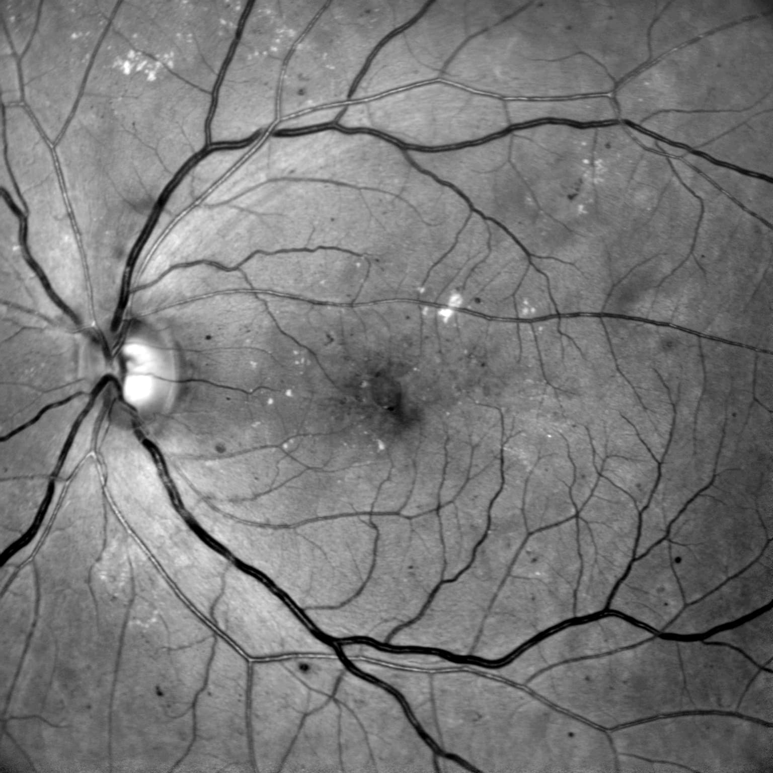
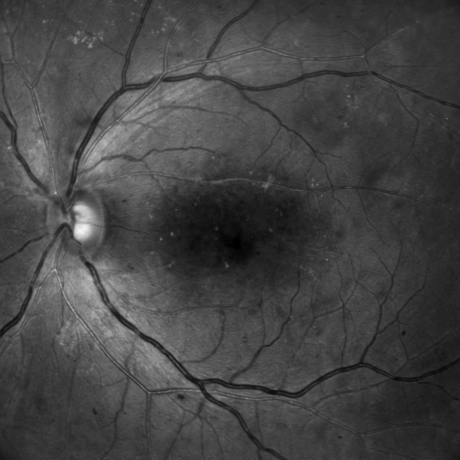
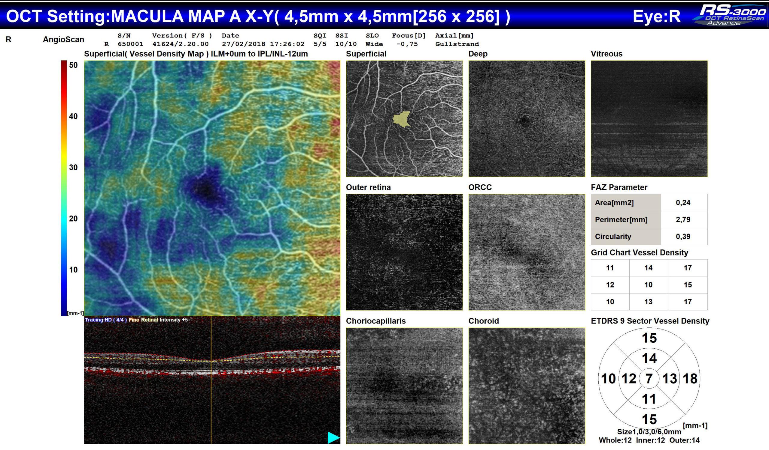
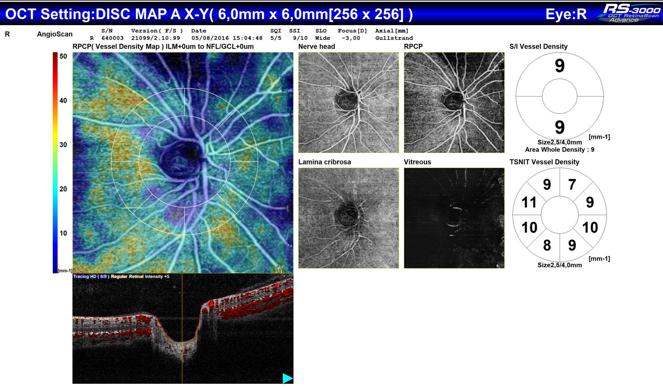
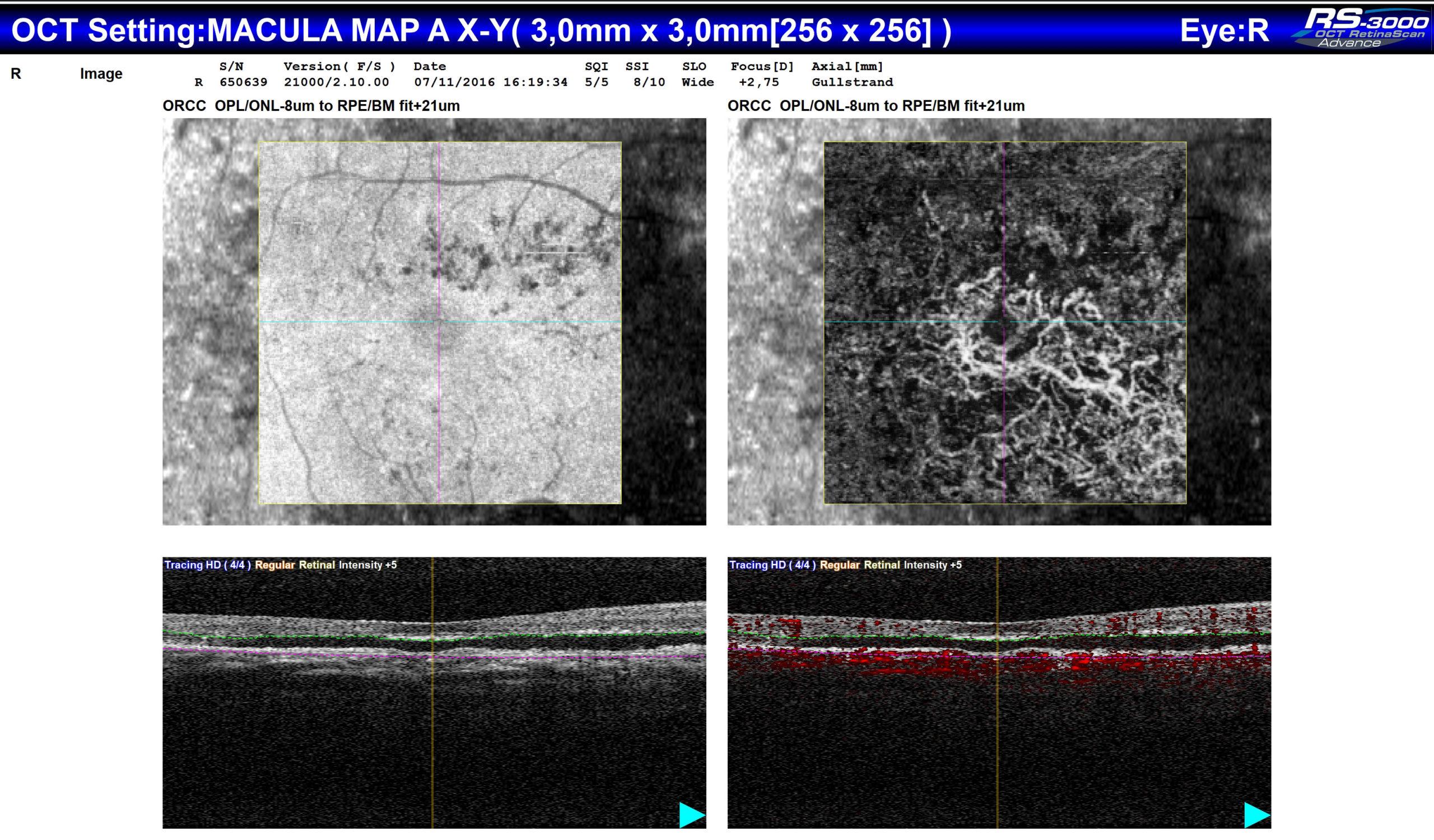
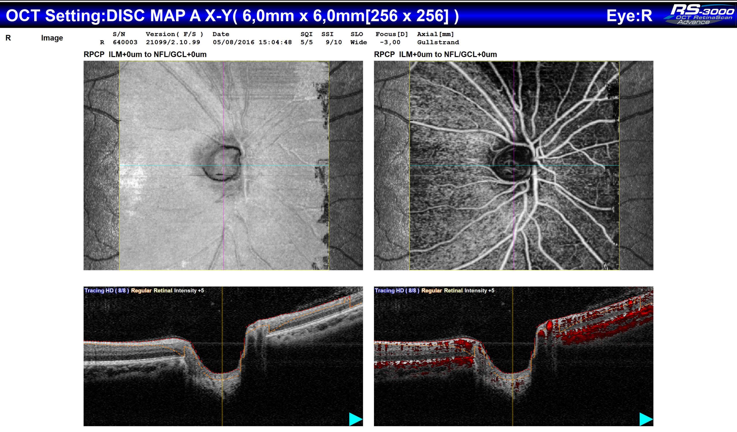
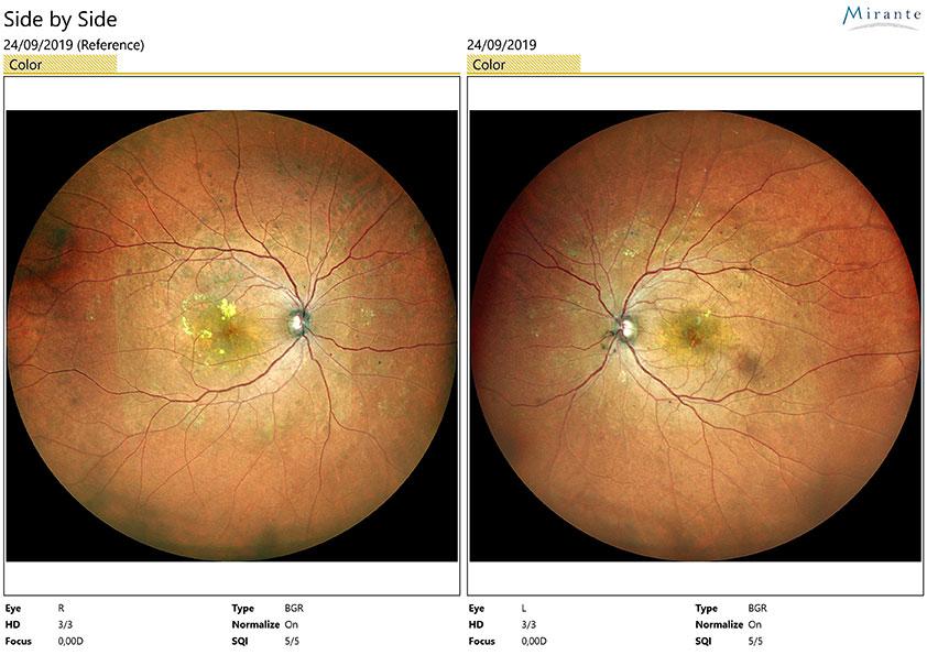
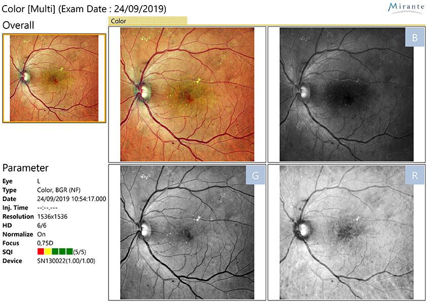
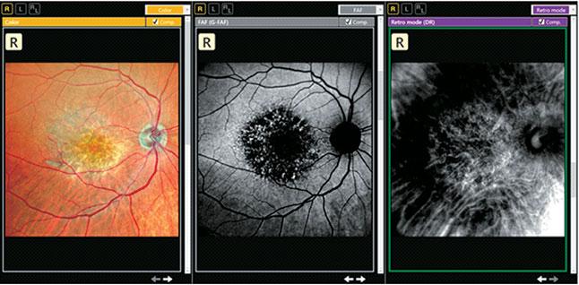
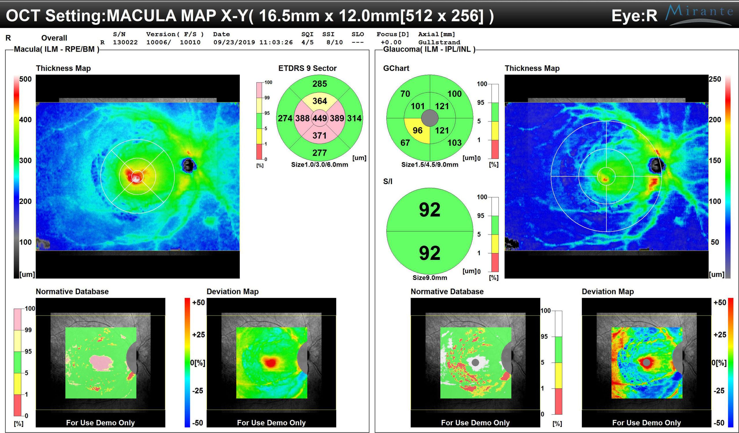
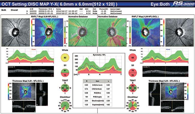
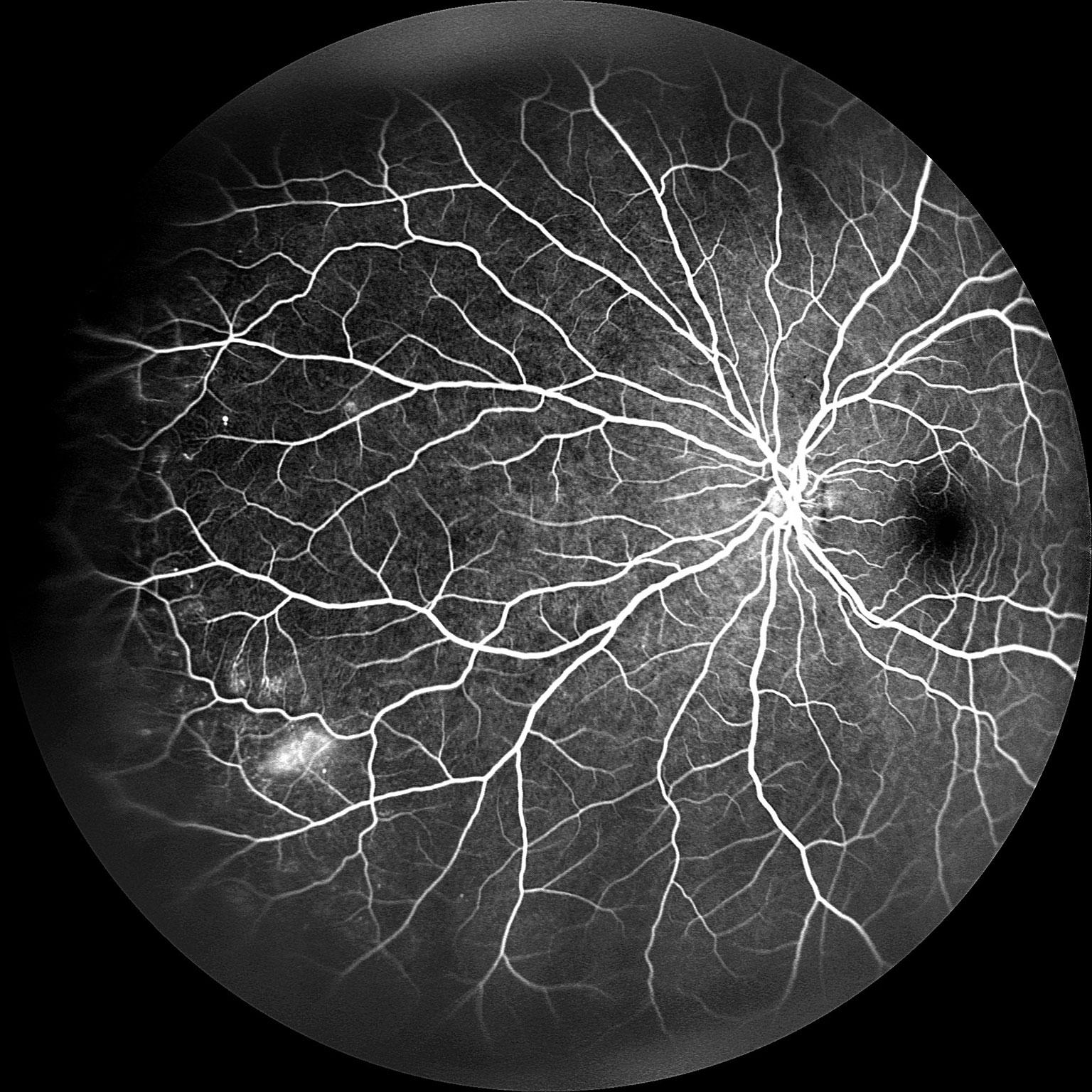
Articles multimodalité :

  • Progress of Imaging in Diabetic Retinopathy—From the Past to the Present – Shintaro Horie and Kyoko Ohno-Matsui
    https://www.ncbi.nlm.nih.gov/pmc/articles/PMC9319818/
  • Assuring Continuity of Retina Care During the COVID-19 Pandemic Using Multimodal Imaging – Aude Couturier, MD, PHD; Ali Erginay, MD; AND Ramin Tadayoni, MD, PHD
  • Multimodal Imaging: Implications for Clinical Practice – BY Giovanni Staurenghi, MD; Srinivas R. Sadda, MD; Mariano Cozzi, MSc; and Giulia Corradetti, MD
  • Widefield Multimodal Imaging – Manish Nagpal, MBBS, MS, FRCS; Gayathri Mohan, MS; Akshar Soni, MS; Sham Talati, DO; and Navneet Mehrotra, MBBS, DNB, FRF
  • Presumed Tenofovir Toxicity – Navneet Mehrotra, MBBS, DNB, FRF, Manish Nagpal, MBBS, MS, FRCS, Sham Talati, DO
  • Comparison between two multimodal imaging platforms: Nidek Mirante and Heidelberg Spectralis – Kimberly Spooner, Long Phan, Mariano Cozzi, Thomas Hong, Giovanni Staurenghi, Eugenia Chu & Andrew A Chang
  • Sensitivity and Specificity of Multimodal Imaging in Characterizing Drusen – Mariano Cozzi, MS; Davide Monteduro, MD; Salvatore Parrulli, MD; Federico Corvi, MD; Federico Zicarelli, MD; Giulia Corradetti, MD; SriniVas R.Sadda, MD; Giovanni Staurenghi, MD


Retours d’expert

  • Utilizing Multimodal & Ultra Wide Field Imaging in the Diagnosis and Management of Retinal Diseases – SriniVas R. Sadda, MD; K. Bailey Freund, MD; and Nadia Waheed, MD
    https://eyetube.net/spotlight/nidek/utilizing-multimodal-ultra-wide-field-imaging-in-the-diagnosis-and-management-of-retinal-diseases
  • 360° Retina Management through Multimodal Imaging: the NIDEK Mirante – Stanislao Rizzo, MD; Thomas Ach, MD; Daniela Bacherini, MD, PhD; and Maria Cristina Savastano, MD, PhD
    https://eyetube.net/spotlight/nidek/360-retina-management-through-multimodal-imaging-the-nidek-mirante
  • The Importance of Multimodal Imaging – Giovanni Staurenghi, MD
  • Multimodal Imaging with the Mirante – Daniela Bacherini, MD, PhD
  • See the Power of Mirante: the Ultimate Multimodal Imaging Platform – Giovanni Staurenghi, MD; SriniVas Sadda, MD; Manish Nagpal, MBBS, MS, FRCS
  • Multimodal & Widefield Imaging – Manish Nagpal, MBBS, MS, FRCS
  • New Horizons in Multimodal Imaging – SriniVas Sadda, MD
  • The importance of multimodal imaging: the new NIDEK Mirante SLO/OCT platform – Giovanni Staurenghi, MD


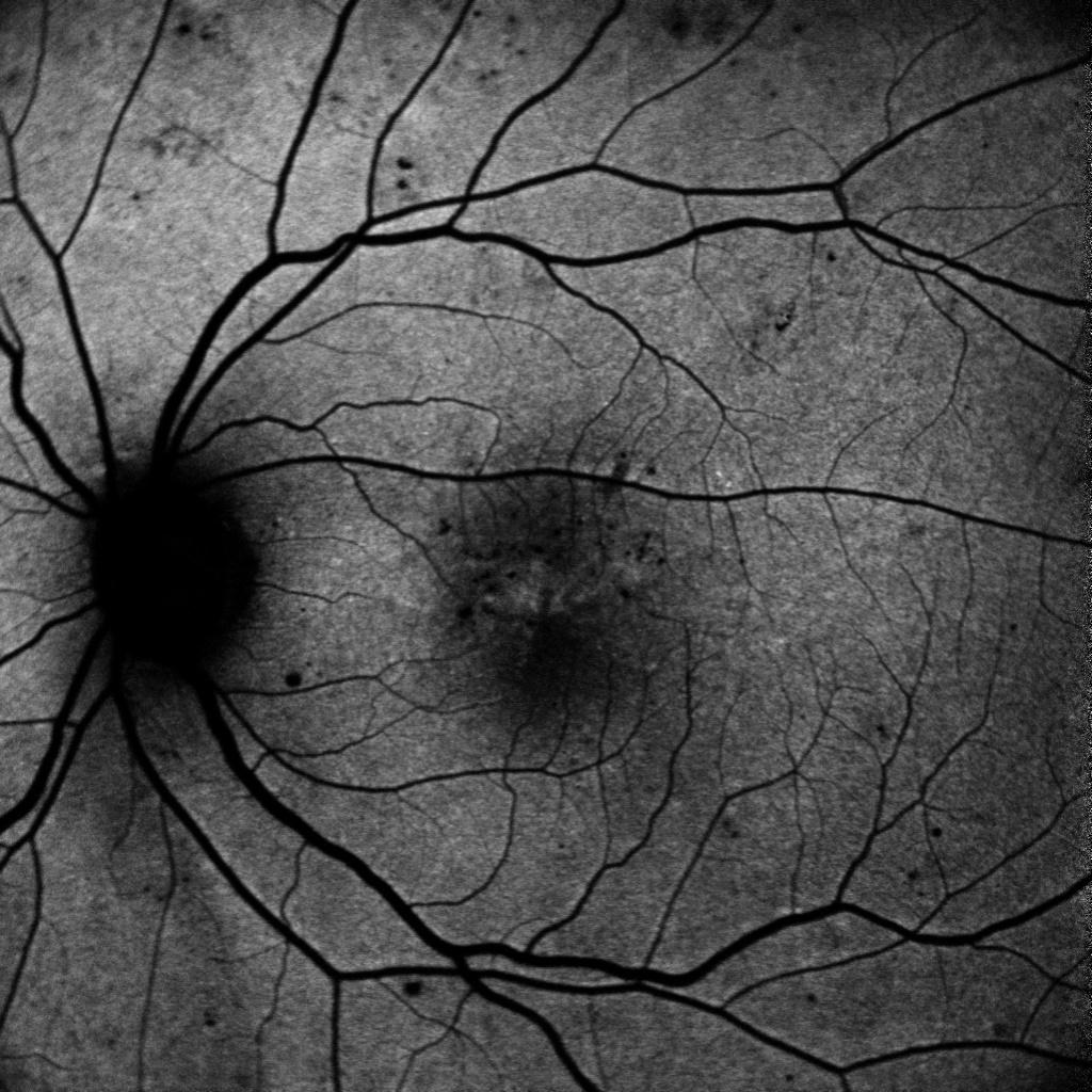
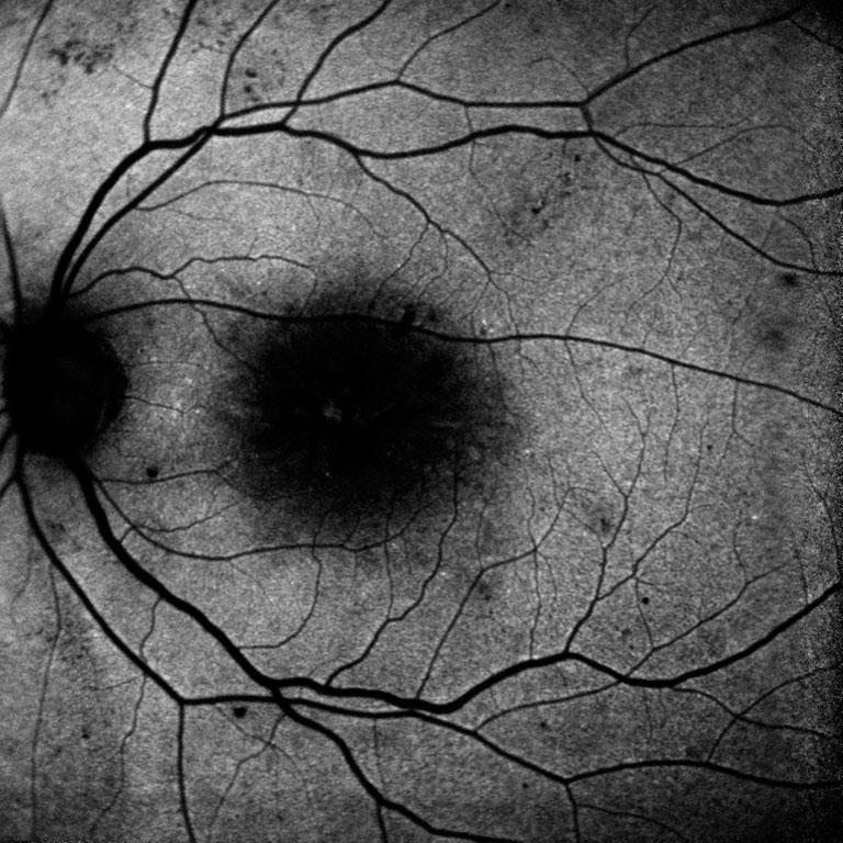
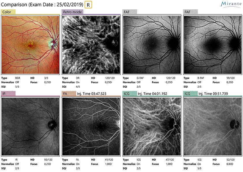
Retours d’expert rétromode :
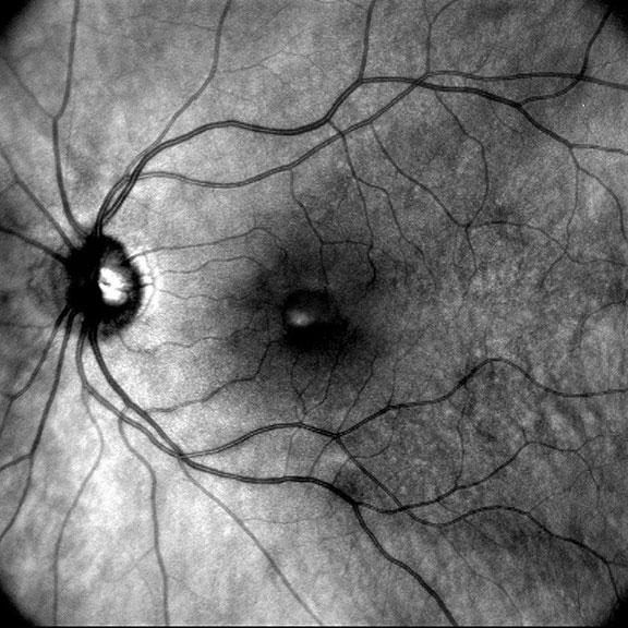
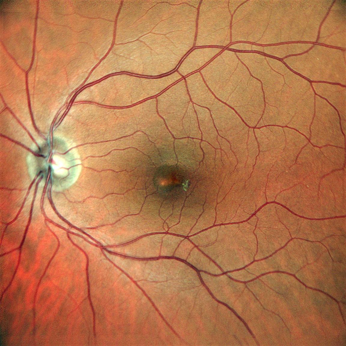
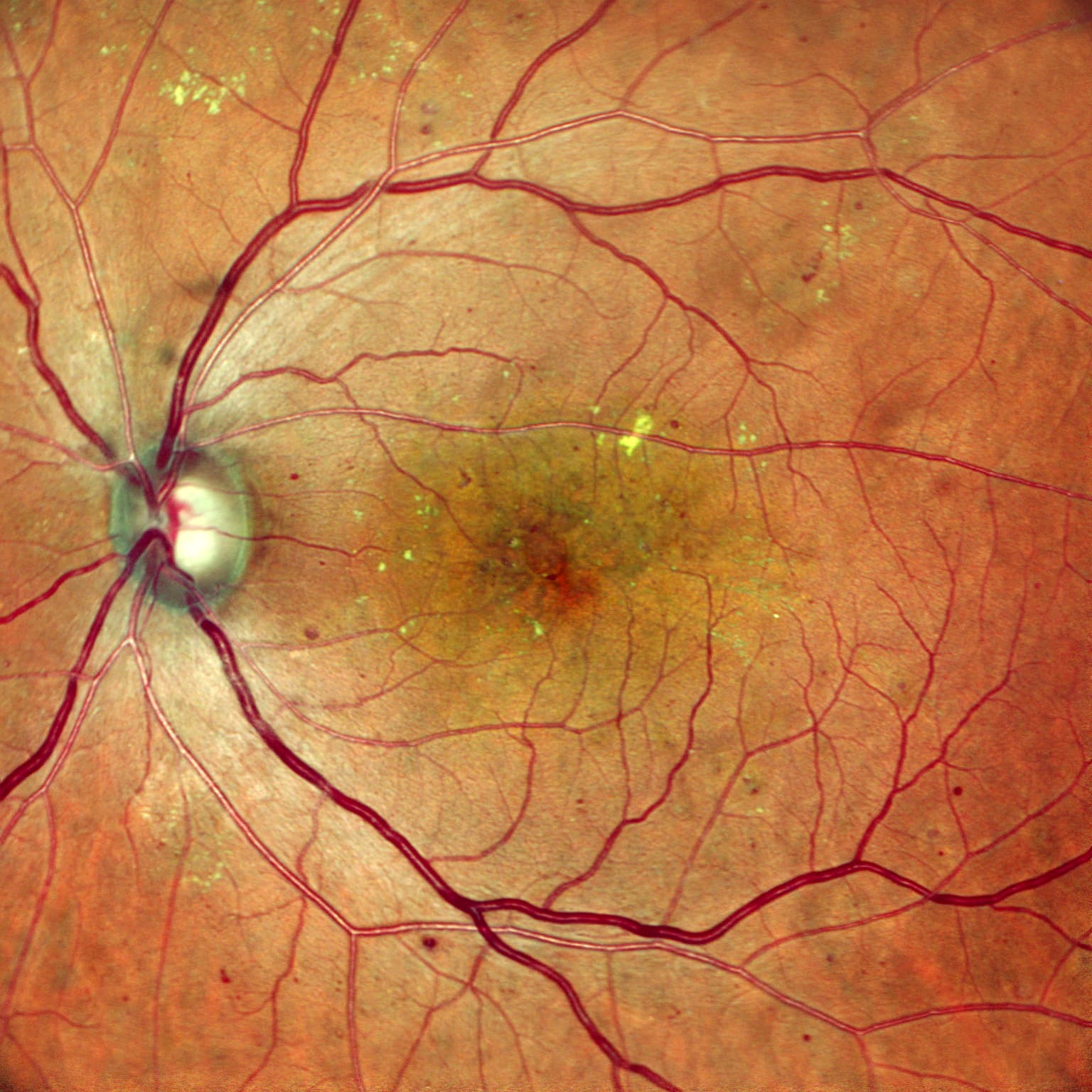
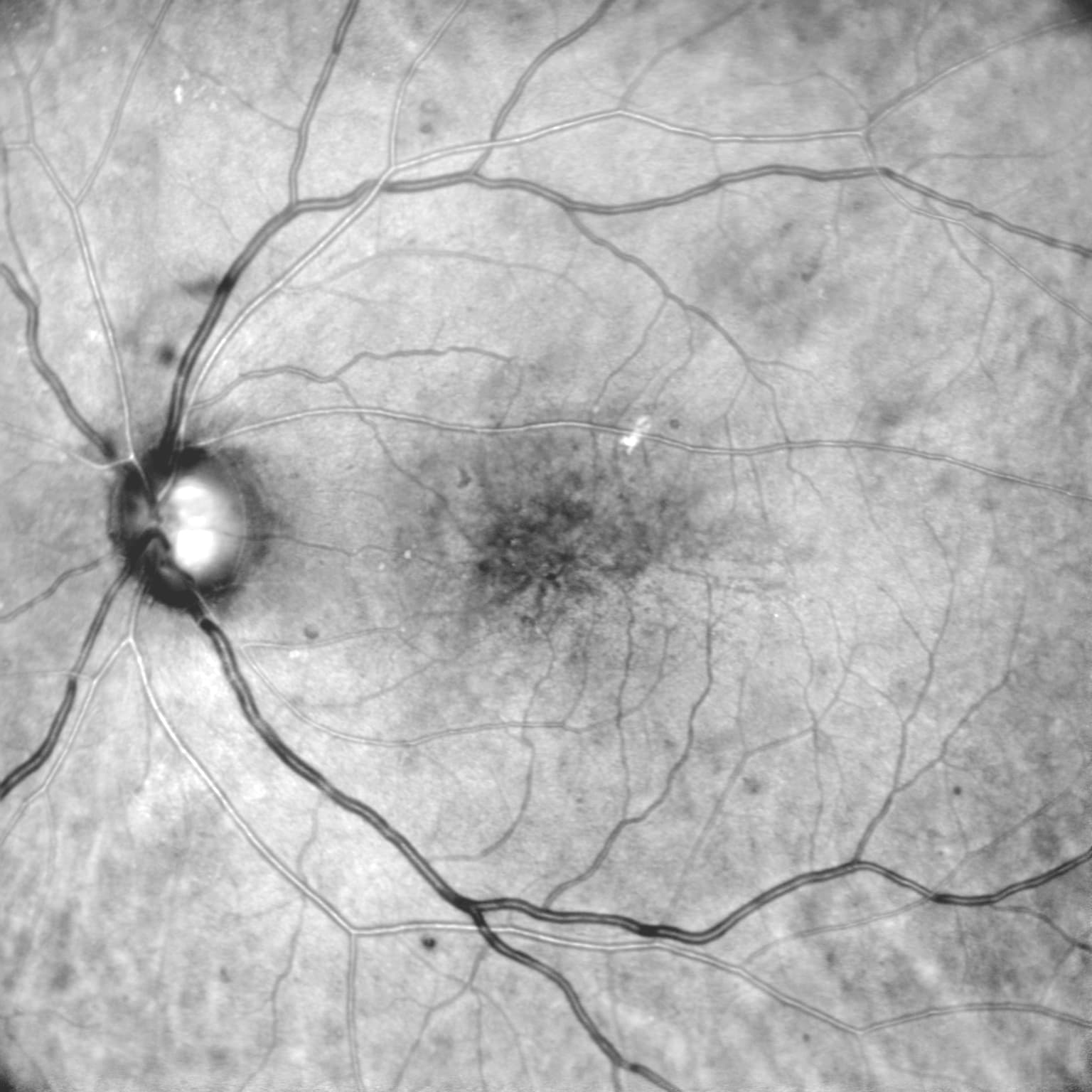
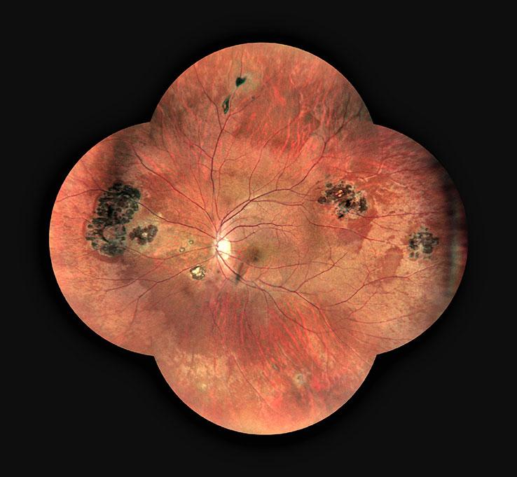
  • Applications of Retro Mode Imaging – SriniVas Sadda, MD
  • Retro Mode Imaging in a Multimodal Assessment of AMD and Macular Dystrophies – SriniVas Sadda, MD
  • Intérêt de l’imagerie rétinienne en RétroMode Prithvi Ramtohul


Présentation de cas

  • SLO-Based Widefield Imaging: An Atlas of Retinal Detachments –

https://retinatoday.com/articles/2022-jan-feb/slo-based-widefield-imaging-an-atlas-of-retinal-detachments?c4src=issue:feed

  • Anti-VEGF Crunch Syndrome in PDR – Yen-Po Chen, MD, PHD

https://retinatoday.com/issues/2022-may-june-insert3

  • Bloody Bowtie, Simultaneous superonasal and superotemporal BRVO – José Gerardo Garcia-Aguirre, MD

https://retinatoday.com/issues/2022-apr-insert2

THE OFFER OF NIDEK PRODUCTS

Additional products to the Mirante Complete Model

Loading...

MP-3

FIND MORE

NAVIS-EX Software

FIND MORE
PRODUCTS YOU MAY ALSO BE INTERESTED IN

In the same range

Loading...

RS-330

FIND MORE

Mirante SLO (SLO)

FIND MORE

Do you require more information?

You have a project? You want a quotation? You have questions about our products?
Feel free to ask your technical sales representative.

Contact us

The art of Eye Care

Reliability and
safety

NIDEK develops its top-of-the-range products to improve visual health through an approach based on strict criteria: safety, reliability, durability, continuous quality controls and certifications.

Technologies and
innovations

NIDEK meets technical challenges by keeping constantly informed of the innovations of eye imaging systems, using the expertise of professionals and the progresses of research.

Services and
guarantees

NIDEK commits itself to providing services to its customers, from the installation of an activity to the authorised training of teams, and to offering long-time measurable guarantees.
FIND MORE

Contact NIDEK

Contact us

Spécialiste des équipements et technologies au service de la santé visuelle, NIDEK SA accompagne et équipe les ophtalmologistes, orthoptistes, professionnels de l’optique et les établissements de santé.

Facebook Linkedin Youtube

KEY PRODUCTS

  • Mirante
  • TS 610
  • RT-Connect
  • Synetic

Services

  • Design office
  • Installation & Implementation
  • Trainings
  • ASS and repairing

Solutions

  • Optics
  • Ophthalmology
  • Orthoptics
  • Health care establishment

About us

  • Expertise and know-how
  • Our key dates
  • Certifications
  • Nidek in France and worldwide
  • Recruitment

© 2021 • NIDEK SA – All rights reserved

  • General Terms and Conditions
  • Protection des données personnelles
  • Legal notices
Gérer le consentement aux cookies
Nous utilisons des cookies pour optimiser notre site web et notre service.
Fonctionnel Always active
Le stockage ou l’accès technique est strictement nécessaire dans la finalité d’intérêt légitime de permettre l’utilisation d’un service spécifique explicitement demandé par l’abonné ou l’utilisateur, ou dans le seul but d’effectuer la transmission d’une communication sur un réseau de communications électroniques.
Préférences
Le stockage ou l’accès technique est nécessaire dans la finalité d’intérêt légitime de stocker des préférences qui ne sont pas demandées par l’abonné ou l’utilisateur.
Statistiques
Le stockage ou l’accès technique qui est utilisé exclusivement à des fins statistiques. Le stockage ou l’accès technique qui est utilisé exclusivement dans des finalités statistiques anonymes. En l’absence d’une assignation à comparaître, d’une conformité volontaire de la part de votre fournisseur d’accès à internet ou d’enregistrements supplémentaires provenant d’une tierce partie, les informations stockées ou extraites à cette seule fin ne peuvent généralement pas être utilisées pour vous identifier.
Marketing
Le stockage ou l’accès technique est nécessaire pour créer des profils d’utilisateurs afin d’envoyer des publicités, ou pour suivre l’utilisateur sur un site web ou sur plusieurs sites web ayant des finalités marketing similaires.
Manage options Manage services Manage {vendor_count} vendors Read more about these purposes
Préférences
{title} {title} {title}

Call me back

A problem was detected in the following Form. Submitting it could result in errors. Please contact the site administrator.

Télécharger votre brochure

Télécharger votre brochure

Télécharger votre brochure

Télécharger votre brochure

Télécharger votre brochure

Télécharger votre brochure

Télécharger votre brochure

Télécharger votre brochure

Télécharger votre brochure

Télécharger votre brochure

Télécharger votre brochure

Télécharger votre brochure

Télécharger votre brochure

Télécharger votre brochure

Télécharger votre brochure

Télécharger votre brochure

Télécharger votre brochure

Télécharger votre brochure

Télécharger votre brochure

Télécharger votre brochure

Télécharger votre brochure

Télécharger votre brochure

Télécharger votre brochure

Télécharger votre brochure

Ce site est réservé aux professionnels de santé

Sortir
Je suis professionnel de santé