Efficient After Sales Service
At your disposal whatever the situation
Made in France
Our consultation units
Our experts at your disposal
60 experts over the territory
Efficient After Sales Service
At your disposal whatever the situation
Made in France
Our consultation units
Our experts at your disposal
60 experts over the territory
Microperimetry is a specific examination used to precisely assess retina sensibility. It can apply to photopic, mesopic or scotopic media; it measures the functionality of cones and rods, which are the retina photoreceptor cells. It completes the detailed morphological analysis provided by the OCT. Once combined, these two methods propose a complete report of the patient’s visual function by correlating retinal structure and function.
Sensitivity analysis aims to determine the extent of lesions caused by certain pathologies, and resulting visual disorders. This examination includes measurement of patient fixation stability, which is important in evaluating vision quality. At the heart of the orthoptist’s work, microperimetry also make it possible to rehabilitate low vision patients suffering from maculopathy and having very low visual acuity.
Unlike the usual examination of the visual field, the microperimetry, which is still a subjective test depending on the patient’s answer, is able to precisely target the analysis zone and to determine the exact location of the visual defect.
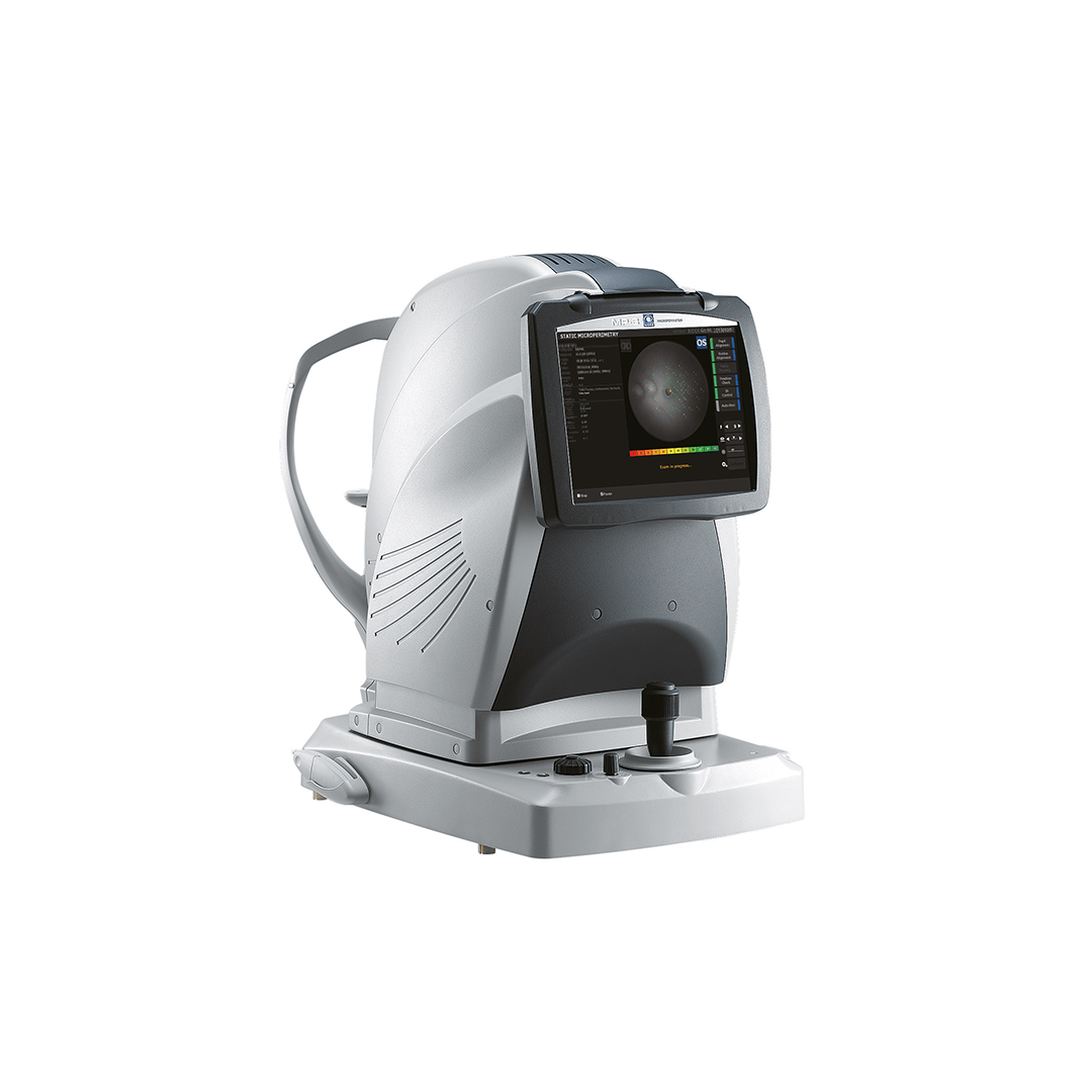
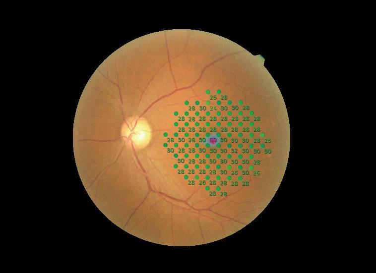
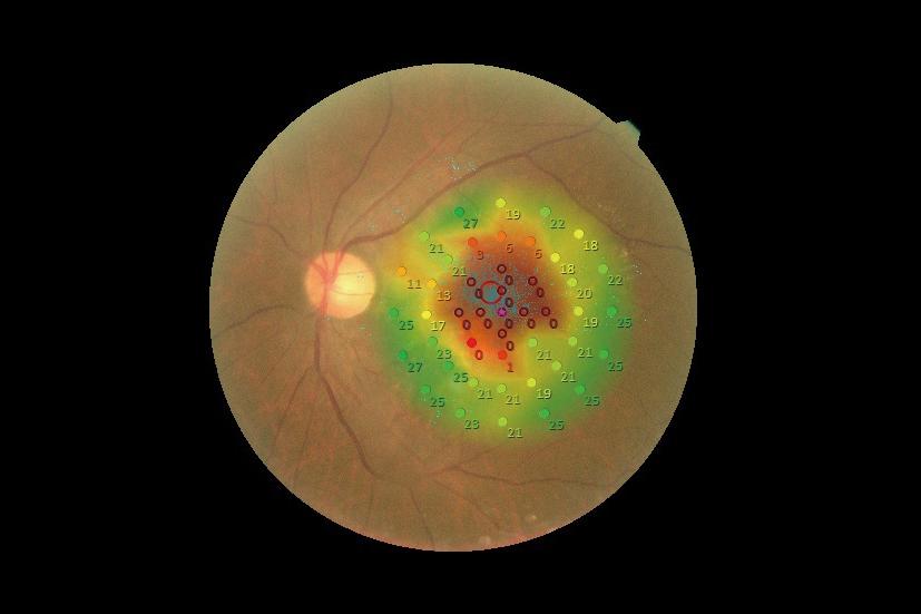
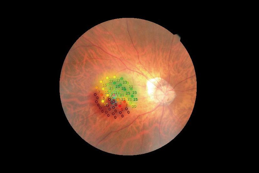
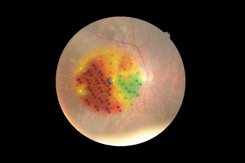
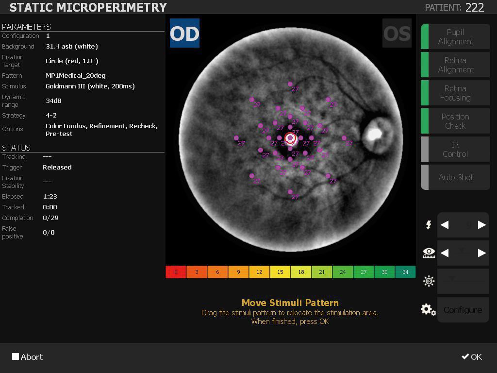
The MP-3S is a microperimeter equipped with a 3D tracking system of the eye and a non-mydriatic fundus camera used to perform microperimetry, to assess the sensibility of photoreceptor cells of the retina. The range of light intensities, from 0 to 34 dB in photopic/mesopic and from 0 to 24 dBin scotopic and the max. luminance of stimuli at 10,000 abs makes possible detecting the lowest sensibilities.
The MP-3S is a microperimeter equipped with a 3D tracking system of the eye and a non-mydriatic fundus camera used to perform microperimetry, to assess the sensibility of photoreceptor cells of the retina. The range of light intensities, from 0 to 34 dB in photopic/mesopic and from 0 to 24 dBin scotopic and the max. luminance of stimuli at 10,000 abs makes possible detecting the lowest sensibilities.
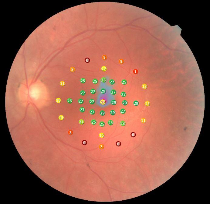
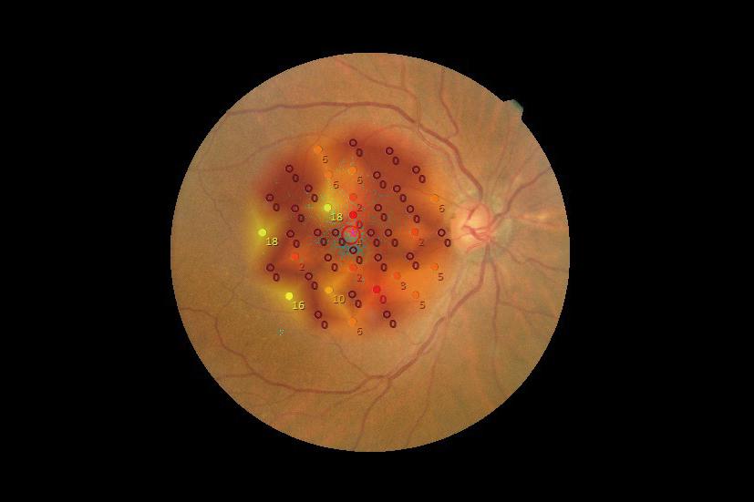
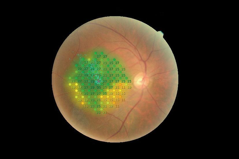
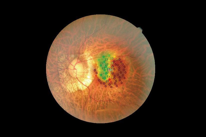
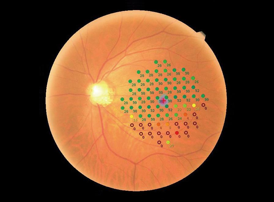
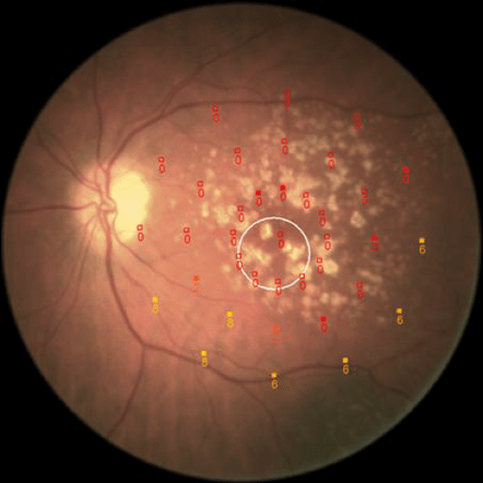
The purpose of microperimetry is assessing the retinal sensibility, the running of the photoreceptor cells of the eye, of the cones and rods, using light stimuli proposed to the patient. The stimuli are emitted with different intensities, with increasing or decreasing values (depending on the strategy selected to determine the threshold) in order to define the response thresholds of retina. Each time the patient detects the stimulus, he/she presses the response bulb, thus indicating to the instrument that he/she has got the intensity of this stimulus. Once the stimulus is not detected any more, the maximal sensibility is set (in dB).
Unlike conventional visual field devices, microperimeters have a three-dimensional retinal tracking system for accurate positioning of stimuli on a selected zone of the retina and therefore the analysis area.
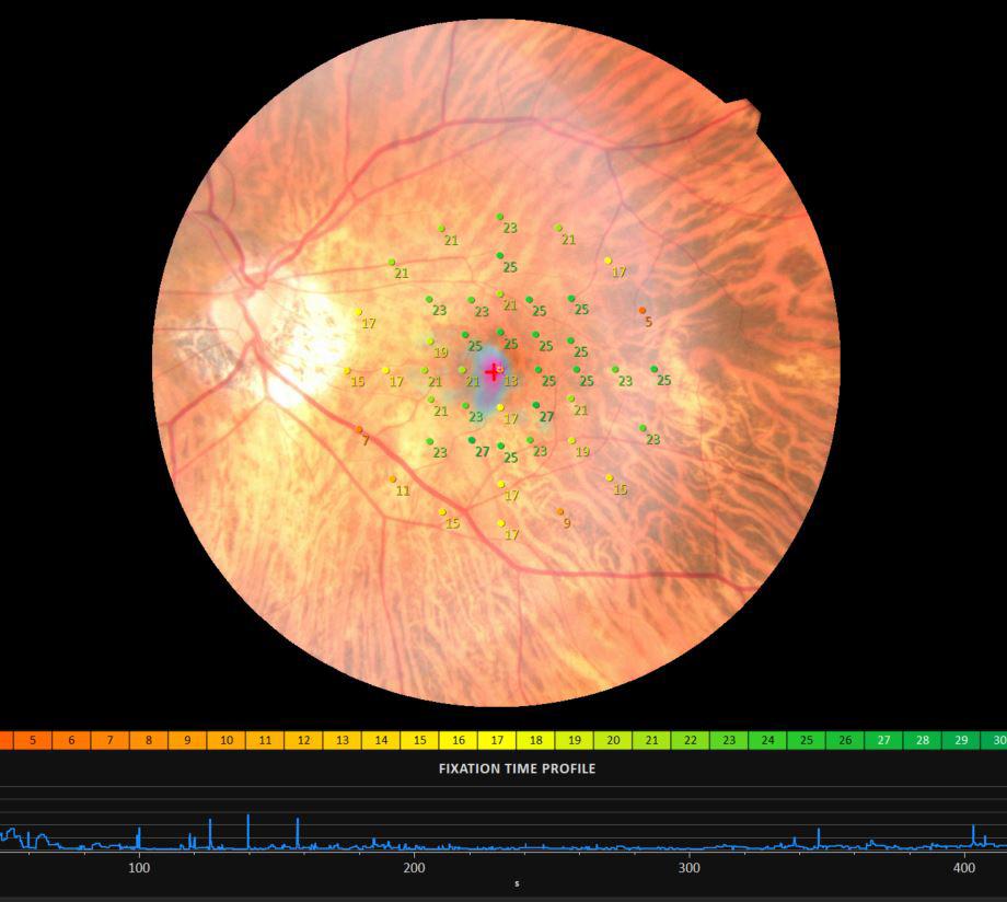
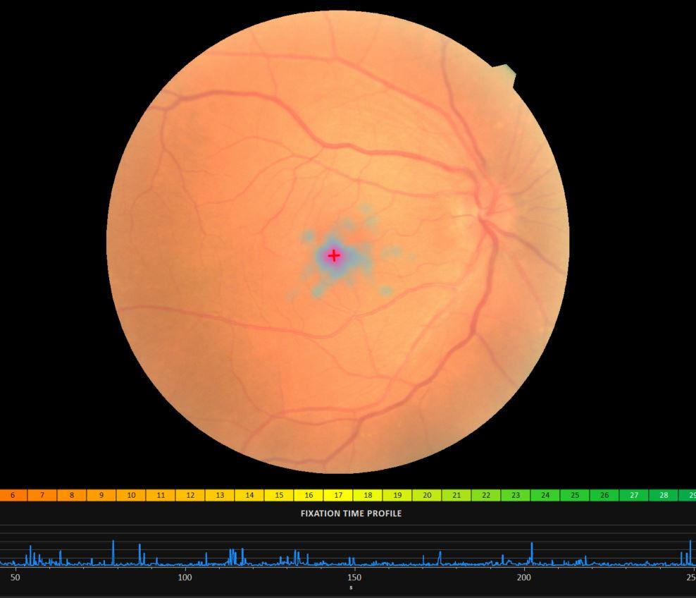
The results are presented on a fundus, in order to view the position of the stimuli and their threshold value. Also, thanks to tracking, the microperimeter follows the patient’s fixation during the microperimetry examination to define fixation stability and evaluate overall visual quality. This information is then used to define visual rehabilitation strategies, primarily in patients with very low visual acuity (low vision).
Daily, any human person continuously tests his environment, particularly by moving his eyes. So, he can stand in the environment and anticipate his motions and moves. Therefore the human eye has a moving strategy, mainly based on central vision.
When a patient has serious visual disturbances, and especially when central vision has been lost, eye movements lose their efficiency and the patient experiences serious difficulties in performing daily tasks.
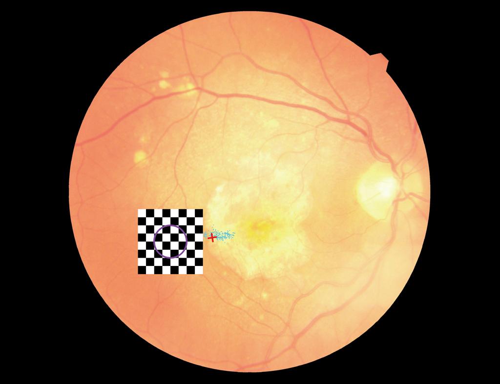
The purpose of rehabilitation, performed using a microperimeter, is training the patient to stabilise his/her fixation and use a better sensibility retina zone to improve the way he/she tests the environment. This training is performed through beeps that guide the patient so he/she can use a better fixation zone.
The microperimetry examination can last several minutes and the patient should concentrate during the whole examination. He/she uses a response switch he/she presses any time he/she detects light stimuli: this is a subjective examination.
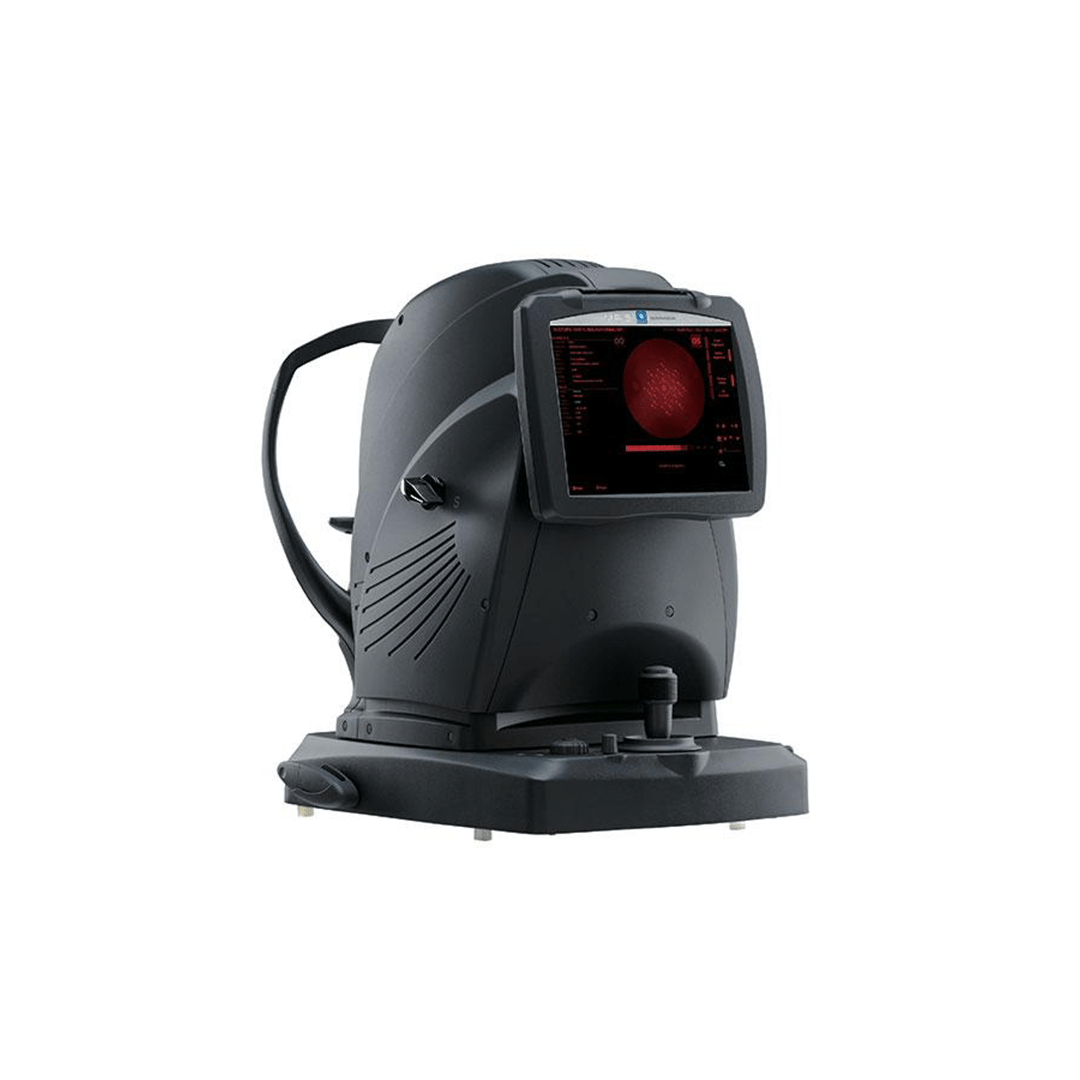
There are two types of photoreceptor cells in charge of vision. As they do not use the same use the same light conditions, there are two types of microperimeter. deux types de micropérimètre.
The scotopic microperimeter can work in the dark, i.e. under night or scotopic light conditions.
The cells that are specifically targeted are rods, which are involved in peripheral vision, contrasts and movements. This exam requires at least 20 minutes of adaptation to darkness.
Microperimetry is the only technique allowing determining the retina sensibility on a precise zone to assess the fixation stability. We can then control the correlation between the structure and the function and better understand the patient’s global visual function.

Equipment with independent eye and retina tracking

Precise positioning of stimuli and overlapping of results on colour photography

Analyses in photopic, mesopic and scotopic conditions

Visual rehabilitation function
You have a project? You want a quotation? You have questions about our products? Feel free to ask your technical sales representative.
Reliability and
safety
NIDEK develops its top-of-the-range products to improve visual health through an approach based on strict criteria: safety, reliability, durability, continuous quality controls and certifications.
Technologies and
innovations
NIDEK meets technical challenges by keeping constantly informed of the innovations of eye imaging systems, using the expertise of professionals and the progresses of research.
Services and
guarantees
NIDEK commits itself to providing services to its customers, from the installation of an activity to the authorised training of teams, and to offering long-time measurable guarantees.
Sucy-en-Brie, le 30 avril 2026 – NIDEK France, leader mondial en équipements d’optique et d’ophtalmologie,
Du 9 au 11 mai 2026, NIDEK vous donne rendez-vous au Palais des Congrès de
NIDEK redéfinît les standards de meulage avec la nouvelle série LEXCE Plus, des meuleuses multifonctions ultra-compactes
1 : Notre bureau d’études pour vous aider sur vos implantations 2 : Espace préconsultation
Du 9 au 11 mai 2026, NIDEK vous donne rendez-vous au Palais des Congrès de
NIDEK SA Lauréate du Mois de l’Économie de la Ville de Saint-Priest Nous sommes heureux
Le Silmo Paris 2025, rendez-vous incontournable de l’optique et de la lunetterie, se tiendra du
Saint-Priest (69), le 12 juin 2025 – NIDEK SA, filiale française du groupe japonais NIDEK,
Ce site est réservé aux professionnels de santé