• 01 49 80 97 97
  • Contact us
  • Boutique NIDEK

Efficient After Sales Service

At your disposal whatever the situation

Made in France

Our consultation units

Our experts at your disposal

60 experts over the territory

  • Lasers
        • Lasers YAG and SLT
          • YC-200 S Plus (YAG/SLT)
          • YC-200 (YAG)
        • Photocoagulator lasers
          • YLC-500 / YLC 500 VIXI
          • GYC-500 / GYC 500 VIXI
  • Retina diagnosis
        • Slit lamps
          • SL-9900/SL-9900 Zoom
          • SL-9800
          • SL-250
        • Multimodal platforms
          • Mirante (SLO/OCT)
          • Mirante SLO (SLO)
        • Fundus cameras
          • AFC-330
          • Retina Scan Duo 2
          • Mirante SLO (SLO)
          • Mirante (SLO/OCT)
        • Microperimeters
          • MP-3
          • MP-3S
        • Angiographs
          • Mirante (SLO/OCT)
          • Mirante SLO (SLO)
        • Software
          • NAVIS-EX Software
        • OCT
          • RS-1
          • Retina Scan Duo 2
          • Mirante (SLO/OCT)
        • Ultrasound scanners
          • US-4000/4000-P (Echo A+B)
          • US-500 / 500-P (Echo A)
  • Anterior segment
        • Slit lamps
          • SL-9900/SL-9900 Zoom
          • SL-9800
          • SL-250
        • Aberrometers - Topographs
          • OPD-Scan III
        • Optical biometers
          • AL-Scan
          • AL-Scan M
        • Tonometers
          • TONOREF III
          • NT-1e / NT-1 / NT-1p
        • Specular microscope
          • CEM-530
        • Goniolenses
          • GS-1
  • Refraction
        • Pupillometers
          • PM-700
        • Afficheurs de tests
          • CP-9
          • SC-1600 Pola/SC-1600
        • Itinerant optician case
          • Itinerant optician case
        • Lensometers
          • LM 1800 P – PD
          • LM7 – 7P
        • Auto refractometers
          • AR-1/ARK-1
          • AR-1a/ARK-1a
          • AR-1s/ARK-1s
          • AR-F/ARK-F
          • HandyRef/HandyRef-K
          • TONOREF III
        • Refractors
          • RT-3100
          • TS-310
          • RT-6100
          • TS-610
          • Logiciel RT-Connect
        • Système de réfraction assisté
          • FARS (Fully Assisted Refraction System)
  • Consultation units
        • "Excellence" units
          • Synetic
          • OT-6400
        • "Performance" units
          • Affinity
          • OT-4200
        • "Compact" units
          • ONE-i
          • Lypop
          • COS-6100
        • Lifting tables and examination swivel chair
          • Consultation armchairs and stools
          • Tables à élévation Up
  • Optical workshop
        • Automatic edges
          • ME-1500
          • LEXCE Trend 8D
          • LEXCE Trend D
          • LEXCE Trend DBT
          • SE9090 Supra
        • Lensometers
          • LM 1800 P – PD
          • LM7 – 7P
        • Centering-blocking devices
          • ICE-1500
          • ICE- 1
        • Shape plotters
          • LT-980
          • LETR-3 Traceur LEXCE Trend
        • Green products
          • LFU-220
          • LED-200 Plus
        • Colonne de centrage : Invision Fit AI
  • NIDEK
        • Our services
          • Design office
          • Installation & Implementation
          • Trainings
          • ASS and repairing
          • Environement
          • Showroom NIDEK
        • Our solutions
          • Optics
          • Ophthalmology
          • Orthoptics
          • Health care establishment
        • About us
          • Expertise and know-how
          • Certifications
          • NIDEK in France
          • NIDEK worldwide
          • Recruitment
        • News
          • Blog
          • Évènements
  • Our shop
  • fr_FR
  • en_GB
PageProduit_MP3_VisuelProduit_Intro

Homepage > Microperimeters > MP-3

MP-3

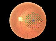
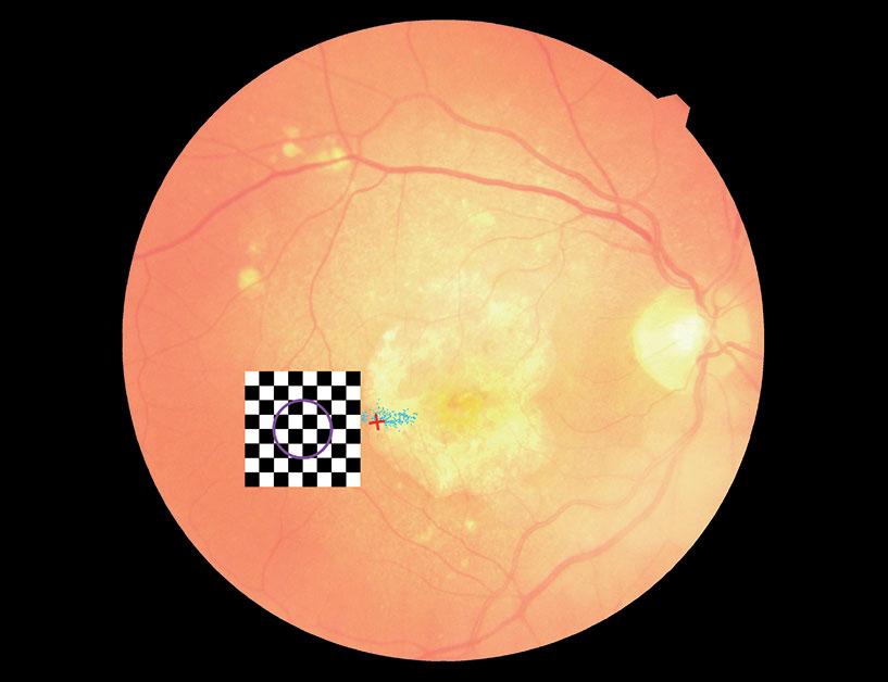
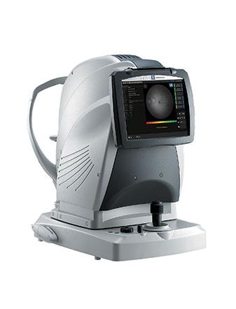
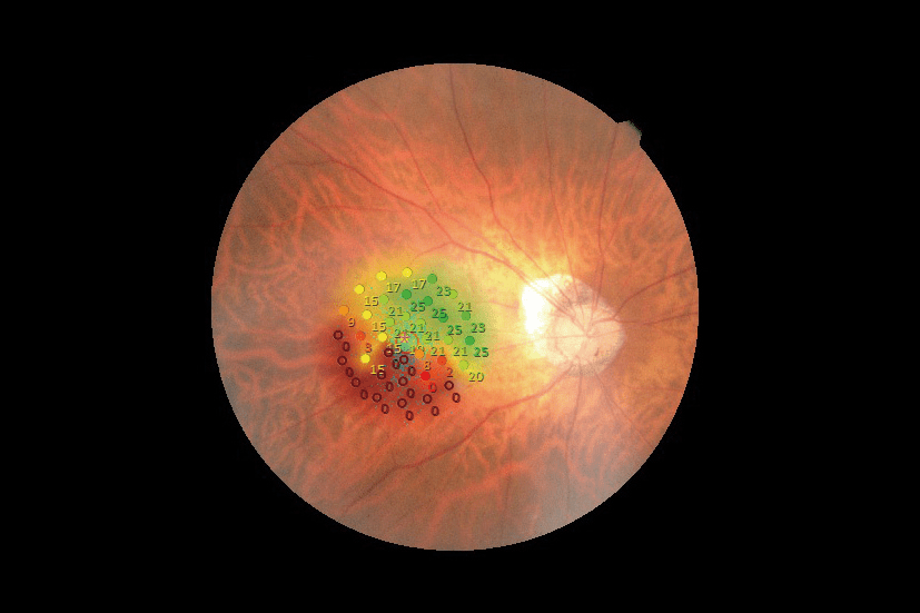
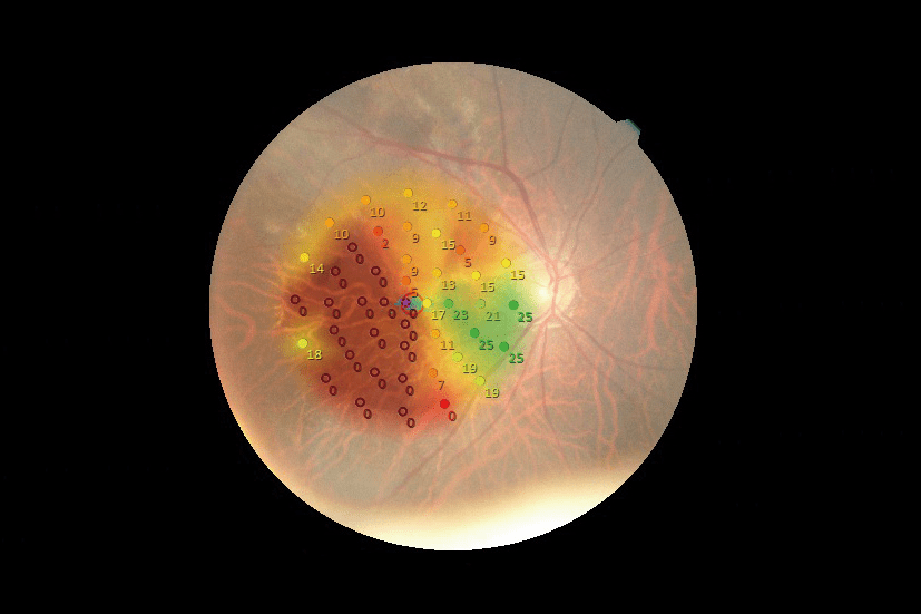
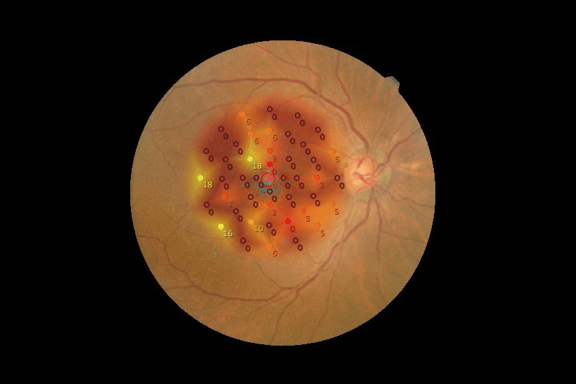
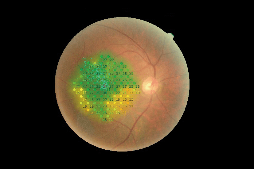
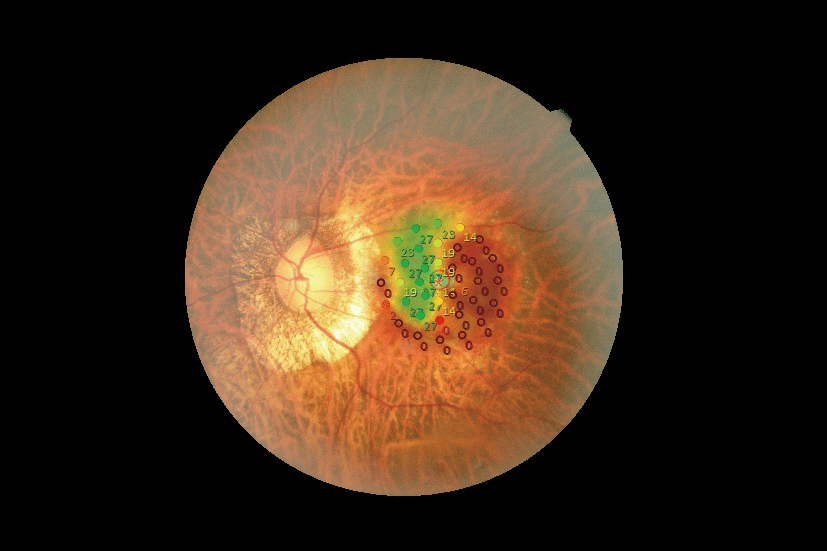
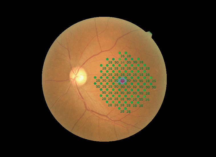
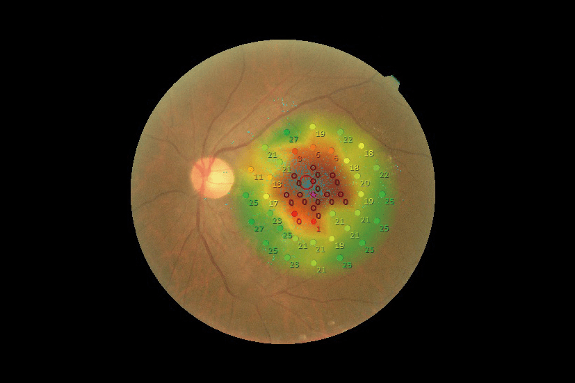
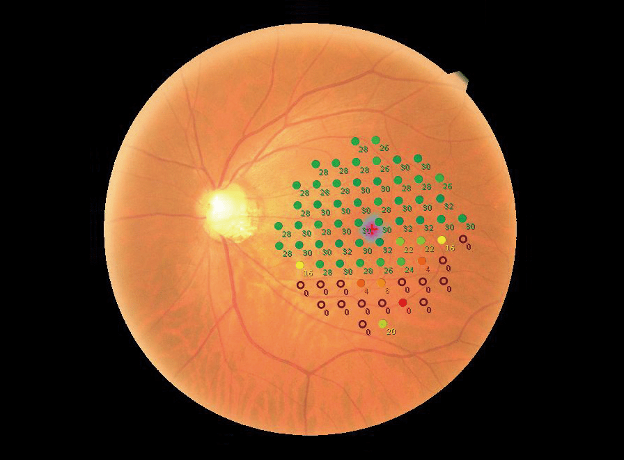
The MP-3 is used to perform the microperimetry exam. It is a very accurate examination used to perform the functional analysis of the retina photoreceptor cells, of the cones and rods, in both photopic and mesopic media. This examination comes with a colour photography of the fundus on which the results superimpose to improve the understanding.

  • More details
  • Get to the gallery
Call me back
Download the brochure
  • Summary
  • Description
  • Accessories
  • Cleaning
  • Documentation
  • Analysis of the sensibility in photopic and mesopic conditions
  • Built-in 12 Mega pixel non-mydriatic fundus camera
  • Real time tracking of the retina and the pupil
  • 40° global analysis field (microperimetry)/45° (retinal)
  • Detection threshold from 0 to 34 dB
  • Stimuli intensity up to 10,000 asb
  • Stimuli gauge in Goldman I, II, III, IV, V
  • Luminance de fond 31,4 asb / 4 asb
  • Dioptrical compensation from -25D to +15D
  • Analysis of the fixation stability
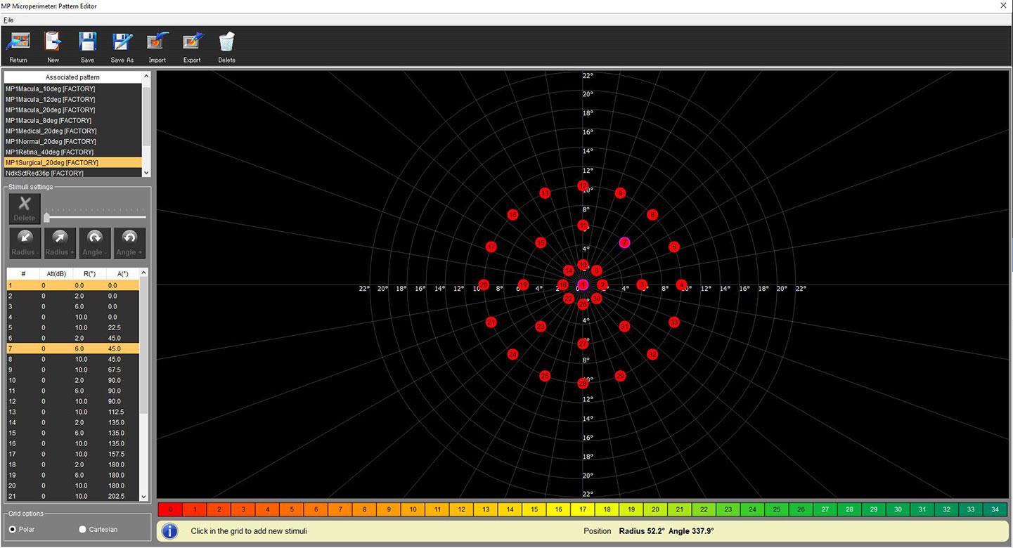
  • Setting the stimuli (stimuli positioning, intensity, etc.) and the fixation point (positioning, size, etc.)
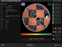
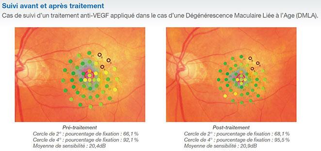
  • Superimposition of the results on the fundus photography
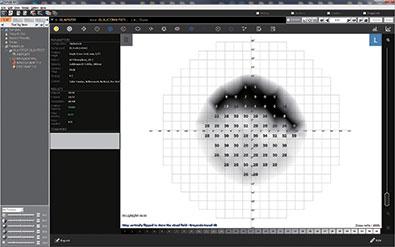
  • Presentation using Humphrey gray scale
  • Feedback function through sound guidance and flickering (variable size grid)
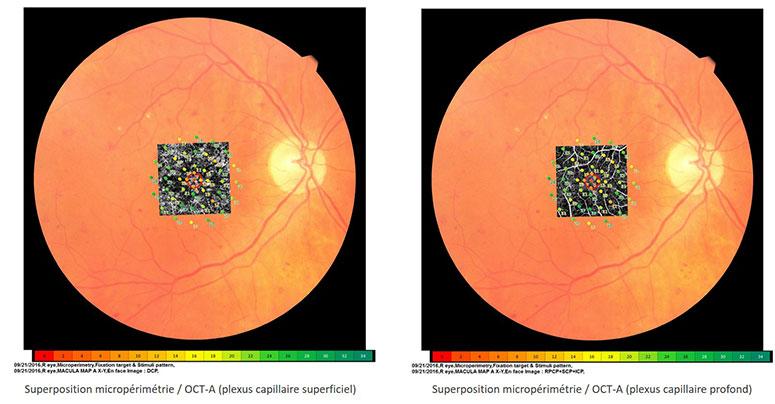
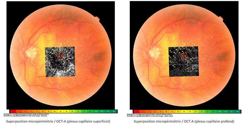
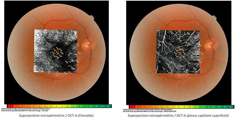
  • Superimposition is possible on OCT/OCT-A

Precise analysis of the visual sensibility and of the fixation

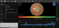
The MP-3 is a microperimeter equipped with a 3D tracking system of the eye and a non-mydriatic fundus camera used to perform microperimetry, to assess the sensibility of photoreceptor cells of the retina. The range of light intensities, from 0 to 34 dB and the max. luminance of stimuli at 10,000 abs makes possible detecting the lowest sensibilities.

This device is equipped with a 3D tracking system of the eye, real time, to correctly position the light stimuli on the retina and limit the patient’s fatigue, as it is quicker. During the examination, the stability of the fixation is also analysed to complete the diagnosis and follow-up the progress of the patient’s rehabilitation. This examination can be performed on its own. It lasts 40 seconds. The analyses are presented either on 2° and 4° circles or in BCEA (Bivariate Contour Ellipse Area).

Résultat de micropérimétrie – Œil glaucomateux
Microperimetry result – Glaucomatous eye
Résultat d’évaluation de la stabilité de fixation
Auto Tracking antérieur et mise au point automatique sur le fond d’œil
Micropérimétrie avec Auto Tracking et Auto alignement
Superposition automatique
Automatic superimposition
Carte différentielle obtenue en Follow-Up
Exemple de choix de rééducation

Patient following-up and rehabilitation

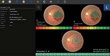
The device itself, thanks to the marks given by the fundus, tracks and aligns to the patient’s eye (anterior and posterior segments) and positions again the results to perform a correct matching with the photography of the fundus. This technology is also used to quantify the evolution of the patients’ sensibility by comparing the similar test zones through a following-up. Then the second examination is automatically carried out in the same conditions as the first one.

The active rehabilitation of the patient (feedback function), mainly used for low-vision patients (with a very low visual acuity), is easily performed, in an optimised way, thanks to the sound guidance (beeps) and the additional visual stimulation (flickering) This procedure reduces the duration of rehabilitation sessions (10 min instead of 30-40 min).

Further analysis

The NAVIS-EX software interface provides an intuitive display of the results and proposes to the user the ability to totally set the examination, particularly using a 100%-adjustable stimuli grid. Thanks to this software, superimposing easily the microperimetry results to the patient OCT exams is possible, to perform a deeper multimodal analysis. To compare the results obtained with the classical visual field devices, a similar Humphrey gray scale is also available.

Legal mentions

These mentions are conforms to the French regulation and may vary depending on circumstances in each country.

Paramétrage des grilles de stimulis
Setting the stimuli grids
Affichage en échelle de gris type Humprhey

Standard accessories

  • External fixation point
  • Response switch
  • Tactile stylus and holder
  • Eye shield
  • Cleaning air gun
  • Protective cover
  • Protective sheet for chinrest
  • Rehabilitation enclosures

Consumables

  • Protective sheet for chinrest
  • Good MP3S cleaning practices
  • Download the brochure


Retours d’expert :

  • Widefield OCT Angiography and Microperimetry in Clinical Practice – Rodrigo Abreu-González, MD, PhD
  • Multimodal Structure and Function Analyisis – Nadia K. Waheed, MD, MPH


Articles :

  • Structural and functional retinal changes in patients with type 2 diabetes without diabetic retinopathy – Qiannan Chai, Yimin Yao, Congrong Guo, Hong Lu, and Jingxue Ma
    https://www.ncbi.nlm.nih.gov/pmc/articles/PMC9258434/


  • Long-term follow-up of retinal morphology and physiology after 2000 mg sildenafil overdose as a means of attempted suicide: a case report – Gen Miura,corresponding author Takayuki Baba, Ryusuke Hashimoto, and Shuichi Yamamoto
    https://www.ncbi.nlm.nih.gov/pmc/articles/PMC9097436/
  • Long-term follow-up of retinal sensitivity assessed by microperimetry in patients with internal limiting membrane peeling – Benjamin Blautain, Agnès Glacet-Bernard, Rocio Blanco-Garavito, Adélaïde Toutée, Camille Jung, Manon Ortoli, Eric H Souied
  • Active Visual Rehabilitation: A New Paradigm in Low Vision Services – Filippo Amore, MD, PhD
  • Structure–Function Correlation Using OCT Angiography And Microperimetry In Diabetic Retinopathy – Alonso-Plasencia M, Abreu-González R, Gómez-Culebras MA
  • Correlation of OCT Angiography and Microperimetry in Wet AMD – Manish Nagpal, MS, DO, FRCS(Edin); Jayesh Khandelwal, MS; Rakesh Juneja, MS; and Navneet Mehrotra, MBBS, DNB (Ophthalmology), FRF
  • Preoperative And Postoperative Macular Sensitivity Assessment Using Microperimetry In Eyes Undergoing Silicone Oil Removal – Nagpal, Manish MS, DO, FRCS; Khandelwal, Jayesh MS; Eltayib, Ahmed MD; Mehrotra, Navneet DNB; Juneja, Rakesh MS
  • Effect of internal limiting membrane peeling on normal retinal function evaluated by microperimetry-3 – Yue Qi, Zengyi Wang, Shi-Ming Li, Qisheng You, Xida Liang, Yanping Yu & Wu Liu
THE OFFER OF NIDEK PRODUCTS

MP3 additional products

Loading...

Mirante (SLO/OCT)

FIND MORE

NAVIS-EX Software

FIND MORE
PRODUCTS YOU MAY ALSO BE INTERESTED IN

In the same range

Loading...

MP-3S

FIND MORE

Do you require more information?

You have a project? You want a quotation? You have questions about our products?
Feel free to ask your technical sales representative.

Contact us

The art of Eye Care

Reliability and
safety

NIDEK develops its top-of-the-range products to improve visual health through an approach based on strict criteria: safety, reliability, durability, continuous quality controls and certifications.

Technologies and
innovations

NIDEK meets technical challenges by keeping constantly informed of the innovations of eye imaging systems, using the expertise of professionals and the progresses of research.

Services and
guarantees

NIDEK commits itself to providing services to its customers, from the installation of an activity to the authorised training of teams, and to offering long-time measurable guarantees.
FIND MORE

Contact NIDEK

Contact us

Spécialiste des équipements et technologies au service de la santé visuelle, NIDEK SA accompagne et équipe les ophtalmologistes, orthoptistes, professionnels de l’optique et les établissements de santé.

Facebook Linkedin Youtube

KEY PRODUCTS

  • Mirante
  • TS 610
  • RT-Connect
  • Synetic

Services

  • Design office
  • Installation & Implementation
  • Trainings
  • ASS and repairing

Solutions

  • Optics
  • Ophthalmology
  • Orthoptics
  • Health care establishment

About us

  • Expertise and know-how
  • Our key dates
  • Certifications
  • Nidek in France and worldwide
  • Recruitment

© 2021 • NIDEK SA – All rights reserved

  • General Terms and Conditions
  • Protection des données personnelles
  • Legal notices
Gérer le consentement aux cookies
Nous utilisons des cookies pour optimiser notre site web et notre service.
Fonctionnel Always active
Le stockage ou l’accès technique est strictement nécessaire dans la finalité d’intérêt légitime de permettre l’utilisation d’un service spécifique explicitement demandé par l’abonné ou l’utilisateur, ou dans le seul but d’effectuer la transmission d’une communication sur un réseau de communications électroniques.
Préférences
Le stockage ou l’accès technique est nécessaire dans la finalité d’intérêt légitime de stocker des préférences qui ne sont pas demandées par l’abonné ou l’utilisateur.
Statistiques
Le stockage ou l’accès technique qui est utilisé exclusivement à des fins statistiques. Le stockage ou l’accès technique qui est utilisé exclusivement dans des finalités statistiques anonymes. En l’absence d’une assignation à comparaître, d’une conformité volontaire de la part de votre fournisseur d’accès à internet ou d’enregistrements supplémentaires provenant d’une tierce partie, les informations stockées ou extraites à cette seule fin ne peuvent généralement pas être utilisées pour vous identifier.
Marketing
Le stockage ou l’accès technique est nécessaire pour créer des profils d’utilisateurs afin d’envoyer des publicités, ou pour suivre l’utilisateur sur un site web ou sur plusieurs sites web ayant des finalités marketing similaires.
Manage options Manage services Manage {vendor_count} vendors Read more about these purposes
Préférences
{title} {title} {title}

Galerie

Suivi de traitement
Superposition micropérimétrie / OCT-A dans le cas d’un œdème maculaire du diabétique
Superposition micropérimétrie / OCT-A dans le cas d’une DMLA
Superposition micropérimétrie / OCT-A dans le cas d’une membrane épi-rétinienne
Epiretinal membrane
Polypoid choroidal vasculopathy
Angiomatous retinal proliferation
Central serous chorioretinopathy
Myopie Forte
Healthy eye
Geographic atrophy (AMD)
Glaucoma

Call me back

A problem was detected in the following Form. Submitting it could result in errors. Please contact the site administrator.

Télécharger votre brochure

Télécharger votre brochure

Télécharger votre brochure

Télécharger votre brochure

Télécharger votre brochure

Télécharger votre brochure

Télécharger votre brochure

Télécharger votre brochure

Télécharger votre brochure

Télécharger votre brochure

Télécharger votre brochure

Télécharger votre brochure

Télécharger votre brochure

Télécharger votre brochure

Télécharger votre brochure

Télécharger votre brochure

Télécharger votre brochure

Télécharger votre brochure

Télécharger votre brochure

Télécharger votre brochure

Télécharger votre brochure

Télécharger votre brochure

Télécharger votre brochure

Télécharger votre brochure

Ce site est réservé aux professionnels de santé

Sortir
Je suis professionnel de santé