• 01 49 80 97 97
  • Contact us
  • Boutique NIDEK

Efficient After Sales Service

At your disposal whatever the situation

Made in France

Our consultation units

Our experts at your disposal

60 experts over the territory

  • Lasers
        • Lasers YAG and SLT
          • YC-200 S Plus (YAG/SLT)
          • YC-200 (YAG)
        • Photocoagulator lasers
          • YLC-500 / YLC 500 VIXI
          • GYC-500 / GYC 500 VIXI
  • Retina diagnosis
        • Slit lamps
          • SL-9900/SL-9900 Zoom
          • SL-9800
          • SL-250
        • Multimodal platforms
          • Mirante (SLO/OCT)
          • Mirante SLO (SLO)
        • Fundus cameras
          • AFC-330
          • Retina Scan Duo 2
          • Mirante SLO (SLO)
          • Mirante (SLO/OCT)
        • Microperimeters
          • MP-3
          • MP-3S
        • Angiographs
          • Mirante (SLO/OCT)
          • Mirante SLO (SLO)
        • Software
          • NAVIS-EX Software
        • OCT
          • RS-1
          • Retina Scan Duo 2
          • Mirante (SLO/OCT)
        • Ultrasound scanners
          • US-4000/4000-P (Echo A+B)
          • US-500 / 500-P (Echo A)
  • Anterior segment
        • Slit lamps
          • SL-9900/SL-9900 Zoom
          • SL-9800
          • SL-250
        • Aberrometers - Topographs
          • OPD-Scan III
        • Optical biometers
          • AL-Scan
          • AL-Scan M
        • Tonometers
          • TONOREF III
          • NT-1e / NT-1 / NT-1p
        • Specular microscope
          • CEM-530
        • Goniolenses
          • GS-1
  • Refraction
        • Pupillometers
          • PM-700
        • Afficheurs de tests
          • CP-9
          • SC-1600 Pola/SC-1600
        • Itinerant optician case
          • Itinerant optician case
        • Lensometers
          • LM 1800 P – PD
          • LM7 – 7P
        • Auto refractometers
          • AR-1/ARK-1
          • AR-1a/ARK-1a
          • AR-1s/ARK-1s
          • AR-F/ARK-F
          • HandyRef/HandyRef-K
          • TONOREF III
        • Refractors
          • RT-3100
          • TS-310
          • RT-6100
          • TS-610
          • Logiciel RT-Connect
        • Système de réfraction assisté
          • FARS (Fully Assisted Refraction System)
  • Consultation units
        • "Excellence" units
          • Synetic
          • OT-6400
        • "Performance" units
          • Affinity
          • OT-4200
        • "Compact" units
          • ONE-i
          • Lypop
          • COS-6100
        • Lifting tables and examination swivel chair
          • Consultation armchairs and stools
          • Tables à élévation Up
  • Optical workshop
        • Automatic edges
          • ME-1500
          • LEXCE Trend 8D
          • LEXCE Trend D
          • LEXCE Trend DBT
          • SE9090 Supra
        • Lensometers
          • LM 1800 P – PD
          • LM7 – 7P
        • Centering-blocking devices
          • ICE-1500
          • ICE- 1
        • Shape plotters
          • LT-980
          • LETR-3 Traceur LEXCE Trend
        • Green products
          • LFU-220
          • LED-200 Plus
        • Colonne de centrage : Invision Fit AI
  • NIDEK
        • Our services
          • Design office
          • Installation & Implementation
          • Trainings
          • ASS and repairing
          • Environement
          • Showroom NIDEK
        • Our solutions
          • Optics
          • Ophthalmology
          • Orthoptics
          • Health care establishment
        • About us
          • Expertise and know-how
          • Certifications
          • NIDEK in France
          • NIDEK worldwide
          • Recruitment
        • News
          • Blog
          • Évènements
  • Our shop
  • fr_FR
  • en_GB

Homepage > Specular microscope

Specular microscope

Anterior segment
Our products
Benefits
Getting equipped
SPECULAR MICROSCOPY

Complete analysis of the corneal endothelium

Since it was first introduced in the 80’s, the endothelial specular microscopy has been playing an essential role in the evaluation and improvement of ophthalmology protocols. NIDEK proposes its solutions for specular microscopy.

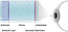
Specular microscopy is used to acquire data and images to perform the cornea assessment. Endothelial cells do not renew and the study of the corneal endothelium (counting of cells and cells quality) makes possible assessing the patient’s situation.

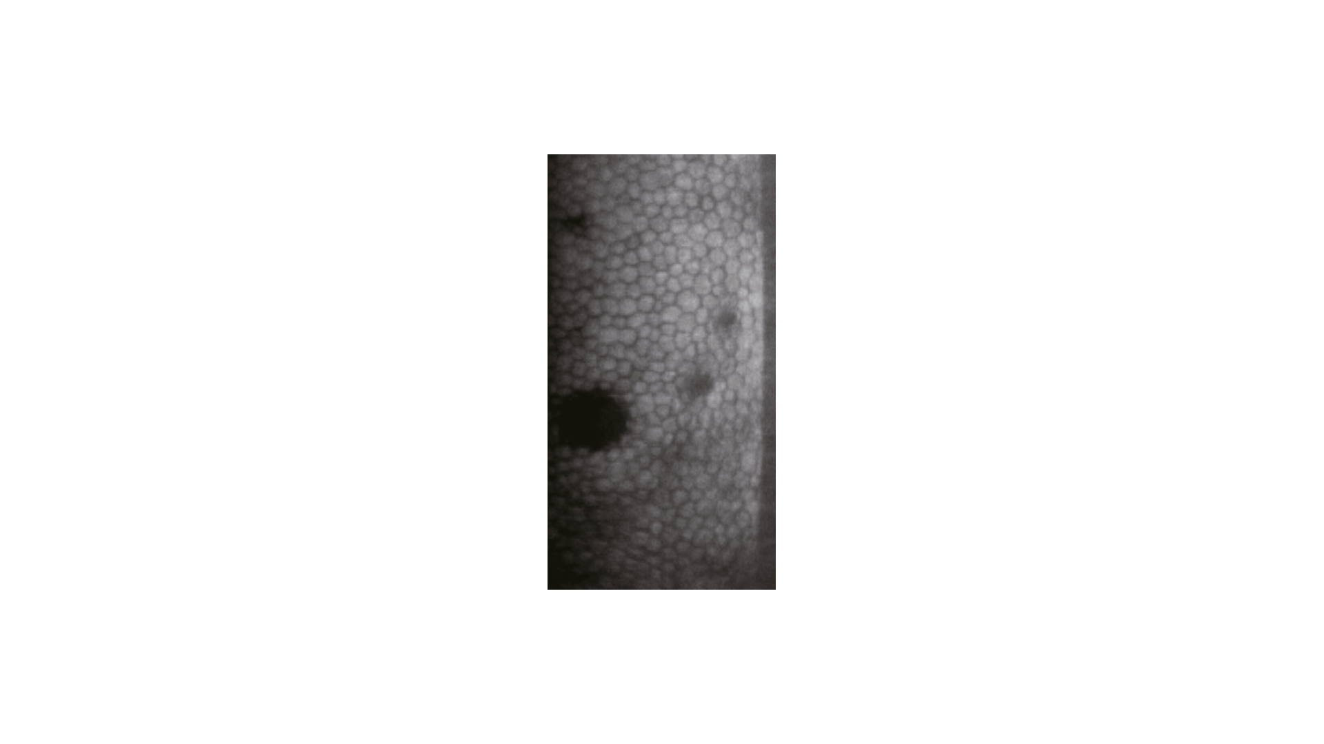
Specular microscopy is a type of examination of endothelial cells. This technique uses the specular reflexion resulting from the interface between the endothelial cells and the aqueous humour. Practically, today, the most frequently used technology is the contactless one, using a large-field specular microscope. Analysing the endothelium specular image consists in evaluating the appearance of cells covering this specific cornea layer to bring out anomalies such as drops or keratic precipitates.

Moreover the operator wants to count the number of endothelial cells in a defined zone while analysing the distribution of the cell sizes. Specular microscopy has been playing a major role in the study of endothelium morphology, and to quantify the endothelial damages produced by different surgery procedures and intraocular systems.

NIDEK PRODUCTS

NIDEK specular microscopes

Loading...

CEM-530

Specular microscope used to perform the automatic acquisition of images and the analysis of corneal endothelium cells without touching the eye.

FIND MORE
Specular microscopy

Techniques used

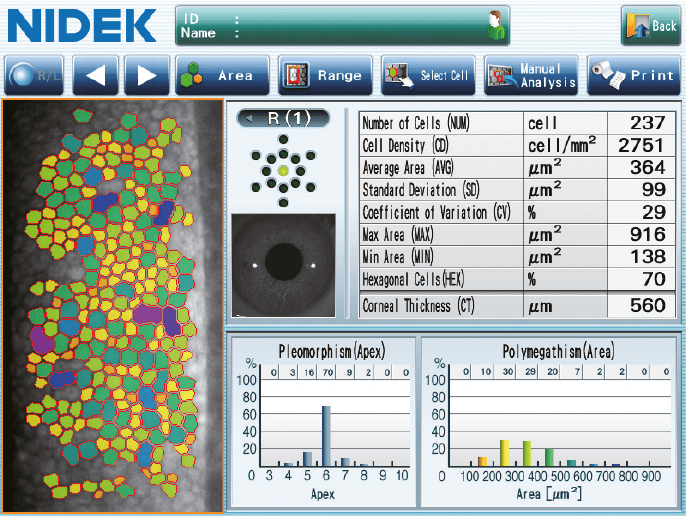
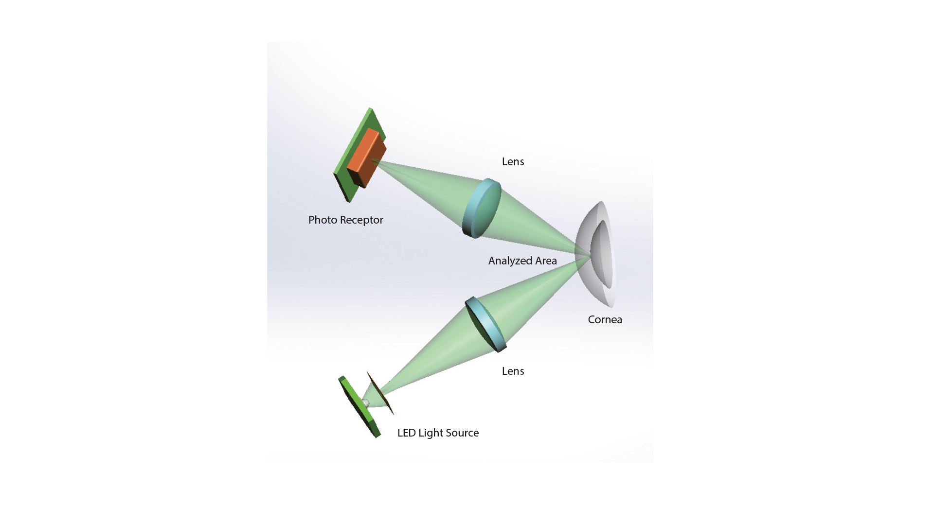
The specular microscope is a non-invasive imaging and analysis device of the corneal endothelial layer. The specular microscopy is an examination of endothelial cells using the specular reflexion resulting from the interface between endothelial cells and aqueous humour.

The specular microscopy can analyse different values, including cell density, size and shape. The technology is based upon a light projection on the cornea posterior face to capture the image reflected by the optical interface between the corneal endothelial layer and the aqueous humour. The image is then automatically analysed and displayed as a specific microphotography.

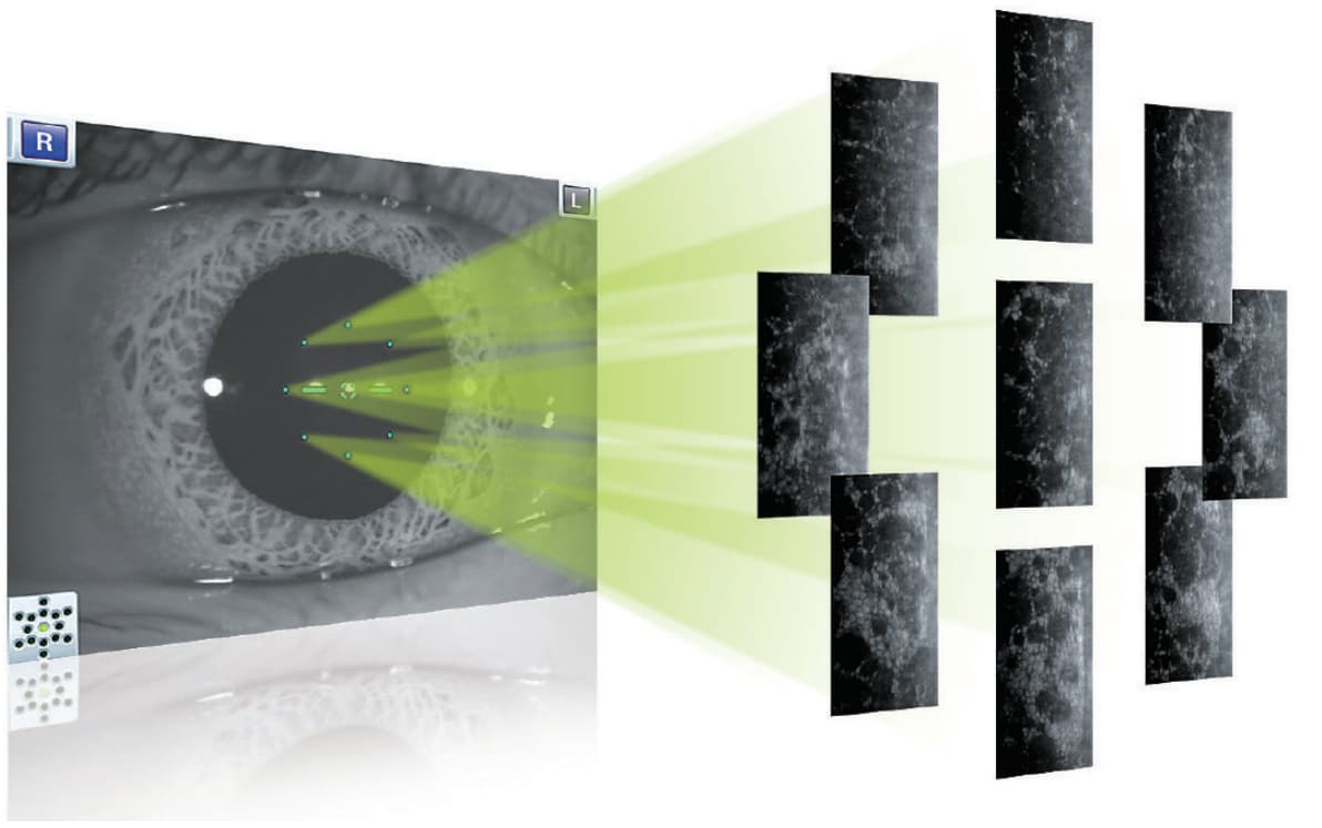
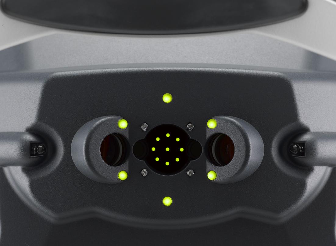
To enable a large investigation zone and a global observation of the cornea, many fixation points are added to central fixation to get images matching both paracentral and peripheral zones.

In clinical practice, specular microscopy if very often used to perform the endothelial examination of cornea. It is also widely used as a pre/post surgery detection system for the cataract surgery. The endothelial layer is examined for the surgery scheduling to prevent complications, such as post surgery corneal clouding.

Pathologies that can be observed by a specular microscope

Analysing the endothelium specular image consists in evaluating the appearance of cells covering this specific cornea layer to bring out anomalies such as drops or keratic precipitates. Very often, the operator has to count the number of cells in a defined area while analysing the distribution of the cell sizes.

Guttata

Fuch's Distrophy

Descemet's Fold Post-DALK

Polymegathism

Images courtesy of Prof. Ekkehard Fabian, Dr. Alaa M. Eldanasoury, Dr. Richard Yudi Hida.
Microscope spéculaire

The different analysis zones

The specular microscope CEM-530 performs central, paracentral (diameter: 1.3 mm) and peripheral (diameter: 7.3 mm) conventional specular microscopies. The combination of central, paracentral and peripheral imaging provides an overview that can be used to make a detailed morphological and quantitative evaluation of the endothelial layer.

Paracentral images

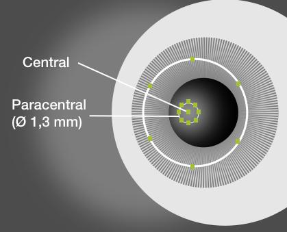
8 target points in paracentral

Paracentral images are captured from 8 points under a visual angle of 5° on a diameter of 1.3 mm to perform an improved evaluation surrounding the central appearance of the cornea.

Peripheral images

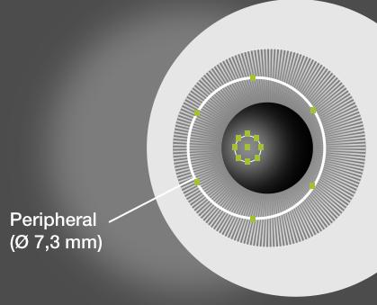
6 target points in periphery

Peripheral images are captured from 6 points under a visual angle of 27° on a diameter of 7.3 mm to perform an improved evaluation of the peripheral zone of the cornea.

Les avantages du microscope spéculaire

The specular microscope CEM-NIDEK is used to rapidly acquire images of endothelium cells and perform a detailed analysis of cell morphology and size.
  • Automation
3D auto-tracking and automatic triggering to improve both accuracy and reproducibility.
  • Exploration

Large investigation zone thanks to target points of central, paracentral and peripheral fixations.

  • Rapidity
Integrated analysis software ensuring the complete analysis of cells, in seconds.

Do you want to purchase a specular microscope?

You have a project? You want a quotation? You have questions about our products? Feel free to ask your technical sales representative.

Contact us

The Art of Eye Care

Reliability and
safety

NIDEK develops its top-of-the-range products to improve visual health through an approach based on strict criteria: safety, reliability, durability, continuous quality controls and certifications.

Technologies and
innovations

NIDEK meets technical challenges by keeping constantly informed of the innovations of eye imaging systems, using the expertise of professionals and the progresses of research.

Services and
guarantees

NIDEK commits itself to providing services to its customers, from the installation of an activity to the authorised training of teams, and to offering long-time measurable guarantees.

FIND MORE
Le magazine

L'actualité NIDEK

Notre actualité

Fermeture exceptionnelle lundi 9 et mardi 10 juin 2025

Chers clients, Nous vous informons qu’exceptionnellement, NIDEK SA sera indisponible les lundi 9 et mardi

Lire la suite »

Les OCT Nidek référencés par l’URPS

Les OCT Nidek référencés par l’URPS : Une opportunité pour les professionnels de santé visuelle

Lire la suite »

🎉 NIDEK à la SFO 2025 : notre gamme médicale à l’honneur !

🎉 NIDEK à la SFO 2025 : notre gamme médicale à l’honneur ! Du 10

Lire la suite »

NIDEK accompagne la formation des assistants médicaux en ophtalmologie avec Keyce Santé

Chez NIDEK, nous sommes convaincus que l’innovation ne se limite pas aux équipements : elle

Lire la suite »
Découvrir le blog
Nos événements

🎉 NIDEK à la SFO 2025 : notre gamme médicale à l’honneur !

🎉 NIDEK à la SFO 2025 : notre gamme médicale à l’honneur ! Du 10

Lire la suite »

Retour sur la visite de M. Ozawa, président de NIDEK, en France

Le 11 février, NIDEK SA France a eu l’honneur d’accueillir M. Ozawa, président de NIDEK,

Lire la suite »

Nidek et MEDADOM renouvellent leur partenariat pour leur dispositif innovant de téléophtalmologie

Communiqué de presse Nidek et MEDADOM renouvellent leur partenariat pour leur dispositif innovant de téléophtalmologie

Lire la suite »

NIDEK SA à la Journée Nationale de Téléophtalmologie 2024

Nous sommes ravis d’annoncer la participation de NIDEK SA à la Journée Nationale de Téléophtalmologie

Lire la suite »
Tous nos événements

Contact NIDEK

Contact us

Spécialiste des équipements et technologies au service de la santé visuelle, NIDEK SA accompagne et équipe les ophtalmologistes, orthoptistes, professionnels de l’optique et les établissements de santé.

Facebook Linkedin Youtube

KEY PRODUCTS

  • Mirante
  • TS 610
  • RT-Connect
  • Synetic

Services

  • Design office
  • Installation & Implementation
  • Trainings
  • ASS and repairing

Solutions

  • Optics
  • Ophthalmology
  • Orthoptics
  • Health care establishment

About us

  • Expertise and know-how
  • Our key dates
  • Certifications
  • Nidek in France and worldwide
  • Recruitment

© 2021 • NIDEK SA – All rights reserved

  • General Terms and Conditions
  • Protection des données personnelles
  • Legal notices
Gérer le consentement aux cookies
Nous utilisons des cookies pour optimiser notre site web et notre service.
Fonctionnel Always active
Le stockage ou l’accès technique est strictement nécessaire dans la finalité d’intérêt légitime de permettre l’utilisation d’un service spécifique explicitement demandé par l’abonné ou l’utilisateur, ou dans le seul but d’effectuer la transmission d’une communication sur un réseau de communications électroniques.
Préférences
Le stockage ou l’accès technique est nécessaire dans la finalité d’intérêt légitime de stocker des préférences qui ne sont pas demandées par l’abonné ou l’utilisateur.
Statistiques
Le stockage ou l’accès technique qui est utilisé exclusivement à des fins statistiques. Le stockage ou l’accès technique qui est utilisé exclusivement dans des finalités statistiques anonymes. En l’absence d’une assignation à comparaître, d’une conformité volontaire de la part de votre fournisseur d’accès à internet ou d’enregistrements supplémentaires provenant d’une tierce partie, les informations stockées ou extraites à cette seule fin ne peuvent généralement pas être utilisées pour vous identifier.
Marketing
Le stockage ou l’accès technique est nécessaire pour créer des profils d’utilisateurs afin d’envoyer des publicités, ou pour suivre l’utilisateur sur un site web ou sur plusieurs sites web ayant des finalités marketing similaires.
Manage options Manage services Manage {vendor_count} vendors Read more about these purposes
Préférences
{title} {title} {title}

Télécharger votre brochure

Télécharger votre brochure

Télécharger votre brochure

Télécharger votre brochure

Télécharger votre brochure

Télécharger votre brochure

Télécharger votre brochure

Télécharger votre brochure

Télécharger votre brochure

Télécharger votre brochure

Télécharger votre brochure

Télécharger votre brochure

Télécharger votre brochure

Télécharger votre brochure

Télécharger votre brochure

Télécharger votre brochure

Télécharger votre brochure

Télécharger votre brochure

Télécharger votre brochure

Télécharger votre brochure

Télécharger votre brochure

Télécharger votre brochure

Télécharger votre brochure

Télécharger votre brochure

Ce site est réservé aux professionnels de santé

Sortir
Je suis professionnel de santé