• 01 49 80 97 97
  • Contact us
  • Boutique NIDEK

Efficient After Sales Service

At your disposal whatever the situation

Made in France

Our consultation units

Our experts at your disposal

60 experts over the territory

  • Lasers
        • Lasers YAG and SLT
          • YC-200 S Plus (YAG/SLT)
          • YC-200 (YAG)
        • Photocoagulator lasers
          • YLC-500 / YLC 500 VIXI
          • GYC-500 / GYC 500 VIXI
  • Retina diagnosis
        • Slit lamps
          • SL-9900/SL-9900 Zoom
          • SL-9800
          • SL-250
        • Multimodal platforms
          • Mirante (SLO/OCT)
          • Mirante SLO (SLO)
        • Fundus cameras
          • AFC-330
          • Retina Scan Duo 2
          • Mirante SLO (SLO)
          • Mirante (SLO/OCT)
        • Microperimeters
          • MP-3
          • MP-3S
        • Angiographs
          • Mirante (SLO/OCT)
          • Mirante SLO (SLO)
        • Software
          • NAVIS-EX Software
        • OCT
          • RS-1
          • Retina Scan Duo 2
          • Mirante (SLO/OCT)
        • Ultrasound scanners
          • US-4000/4000-P (Echo A+B)
          • US-500 / 500-P (Echo A)
  • Anterior segment
        • Slit lamps
          • SL-9900/SL-9900 Zoom
          • SL-9800
          • SL-250
        • Aberrometers - Topographs
          • OPD-Scan III
        • Optical biometers
          • AL-Scan
          • AL-Scan M
        • Tonometers
          • TONOREF III
          • NT-1e / NT-1 / NT-1p
        • Specular microscope
          • CEM-530
        • Goniolenses
          • GS-1
  • Refraction
        • Pupillometers
          • PM-700
        • Afficheurs de tests
          • CP-9
          • SC-1600 Pola/SC-1600
        • Itinerant optician case
          • Itinerant optician case
        • Lensometers
          • LM 1800 P – PD
          • LM7 – 7P
        • Auto refractometers
          • AR-1/ARK-1
          • AR-1a/ARK-1a
          • AR-1s/ARK-1s
          • AR-F/ARK-F
          • HandyRef/HandyRef-K
          • TONOREF III
        • Refractors
          • RT-3100
          • TS-310
          • RT-6100
          • TS-610
          • Logiciel RT-Connect
        • Système de réfraction assisté
          • FARS (Fully Assisted Refraction System)
  • Consultation units
        • "Excellence" units
          • Synetic
          • OT-6400
        • "Performance" units
          • Affinity
          • OT-4200
        • "Compact" units
          • ONE-i
          • Lypop
          • COS-6100
        • Lifting tables and examination swivel chair
          • Consultation armchairs and stools
          • Tables à élévation Up
  • Optical workshop
        • Automatic edges
          • ME-1500
          • LEXCE Trend 8D
          • LEXCE Trend D
          • LEXCE Trend DBT
          • SE9090 Supra
        • Lensometers
          • LM 1800 P – PD
          • LM7 – 7P
        • Centering-blocking devices
          • ICE-1500
          • ICE- 1
        • Shape plotters
          • LT-980
          • LETR-3 Traceur LEXCE Trend
        • Green products
          • LFU-220
          • LED-200 Plus
        • Colonne de centrage : Invision Fit AI
  • NIDEK
        • Our services
          • Design office
          • Installation & Implementation
          • Trainings
          • ASS and repairing
          • Environement
          • Showroom NIDEK
        • Our solutions
          • Optics
          • Ophthalmology
          • Orthoptics
          • Health care establishment
        • About us
          • Expertise and know-how
          • Certifications
          • NIDEK in France
          • NIDEK worldwide
          • Recruitment
        • News
          • Blog
          • Évènements
  • Our shop
  • fr_FR
  • en_GB
RS-3000Advance2_right copie

Homepage > OCT > RS-3000 Advance 2

RS-3000 Advance 2

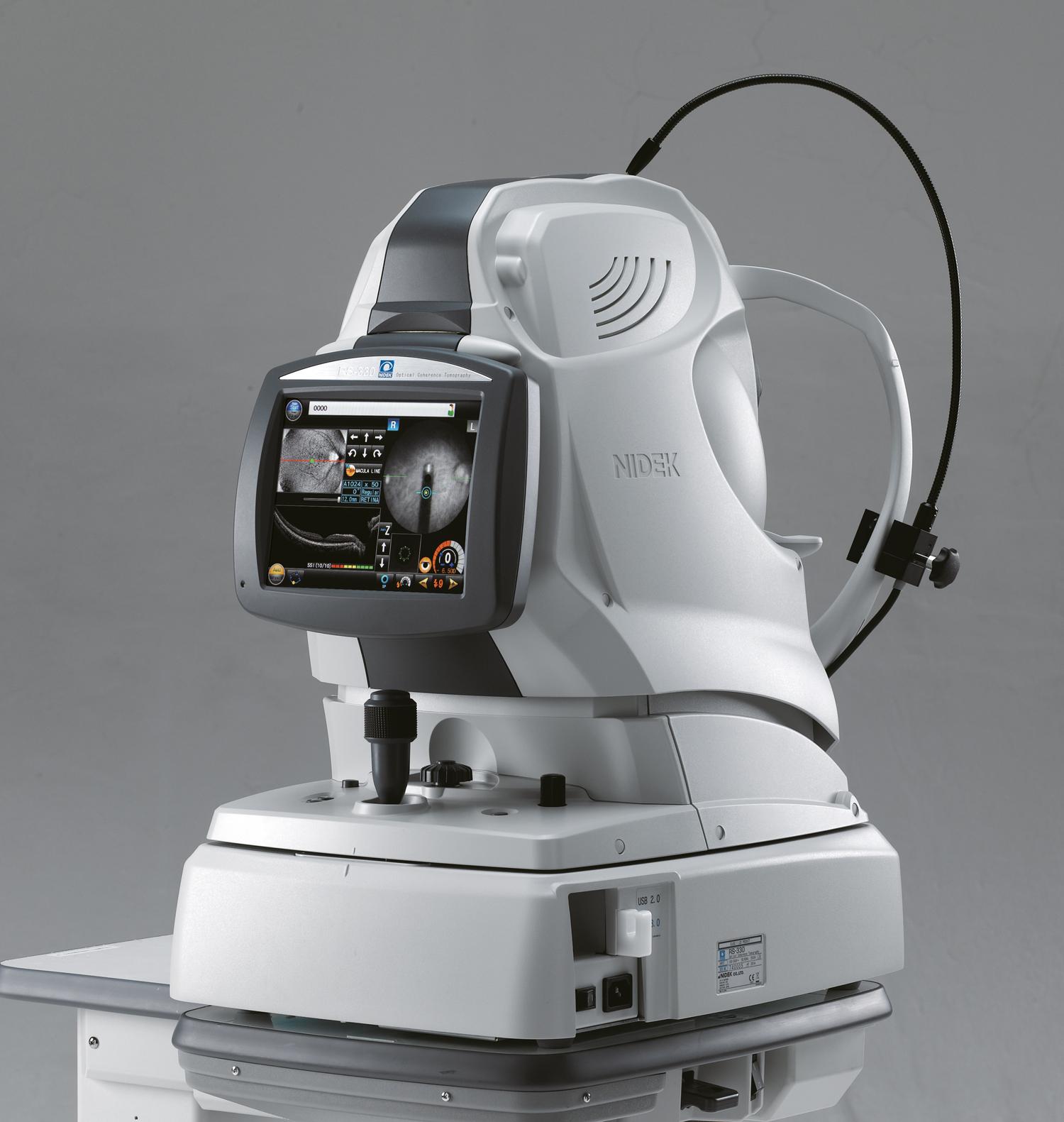
The RS-3000 Advance 2 is an OCT Spectral Domain Expert equipped with technology SLO and a function OCT-Angiography. These advanced functions make it a powerful diagnosis tool of retinal-choroidal pathologies. Thanks to an additional lens, it also makes possible analysing sections of the anterior segment.

  • More details
  • Get to the gallery
Call me back
Download the brochure
  • Summary
  • Description
  • Accessories
  • Cleaning
  • Documentation
  • OCT Expert SLO SD à 85 000 A-scans/s
  • AngioScan module for OCT-Angiography
  • Module OCT B-Scan Denoising
  • LSO retina tracking:
    • Taking into account cyclotorsion
    • HD mode up to 120 sections
    • Real time automatic repositioning of scans
    • Follow-up upon acquisition
    • Accurate OCT-Angiography
  • EDI mode for the choroid
  • Easy to use: automatic focus and positioning of the scans
  • COMBO mode to delegate the tasks
  • Initial dioptrical compensation from -15D to +10D
  • Modulation of the signal sensibility for opaque media
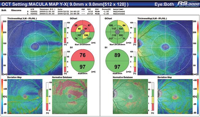
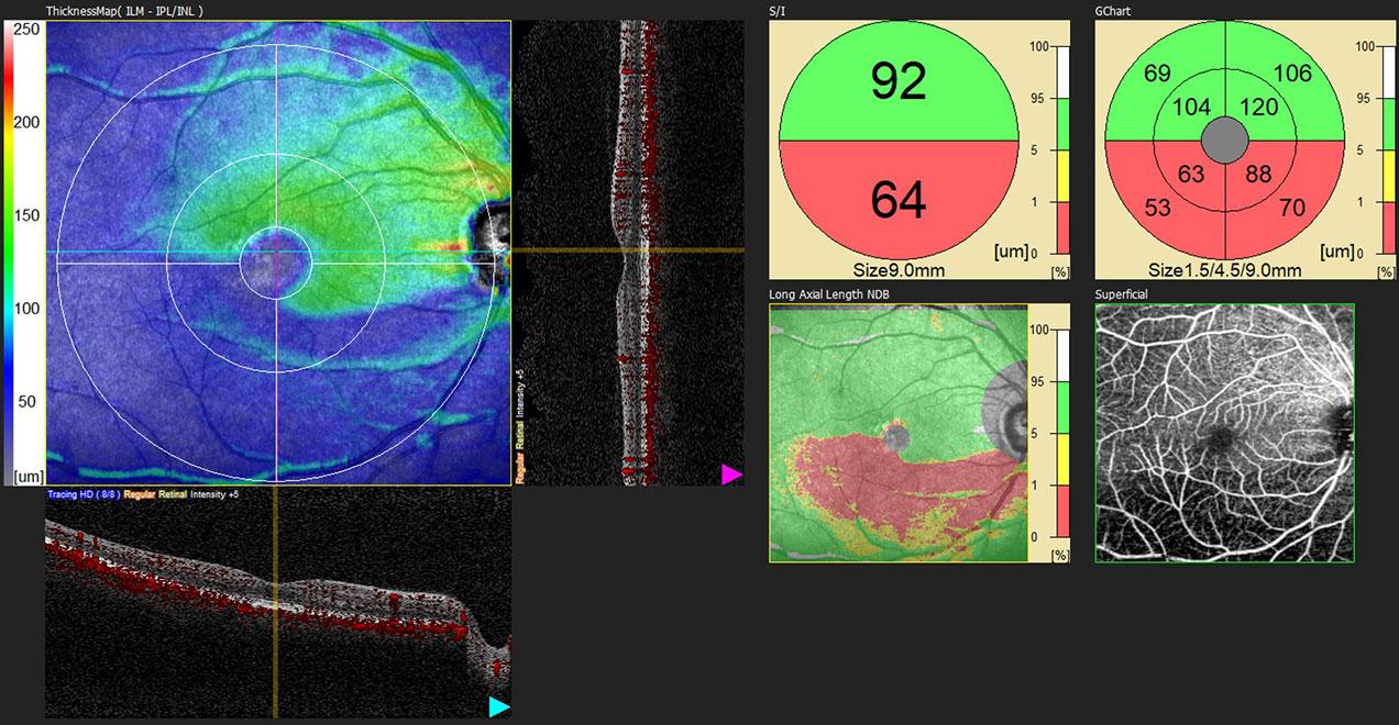
  • Macular analysis with complex of ganglionic cells
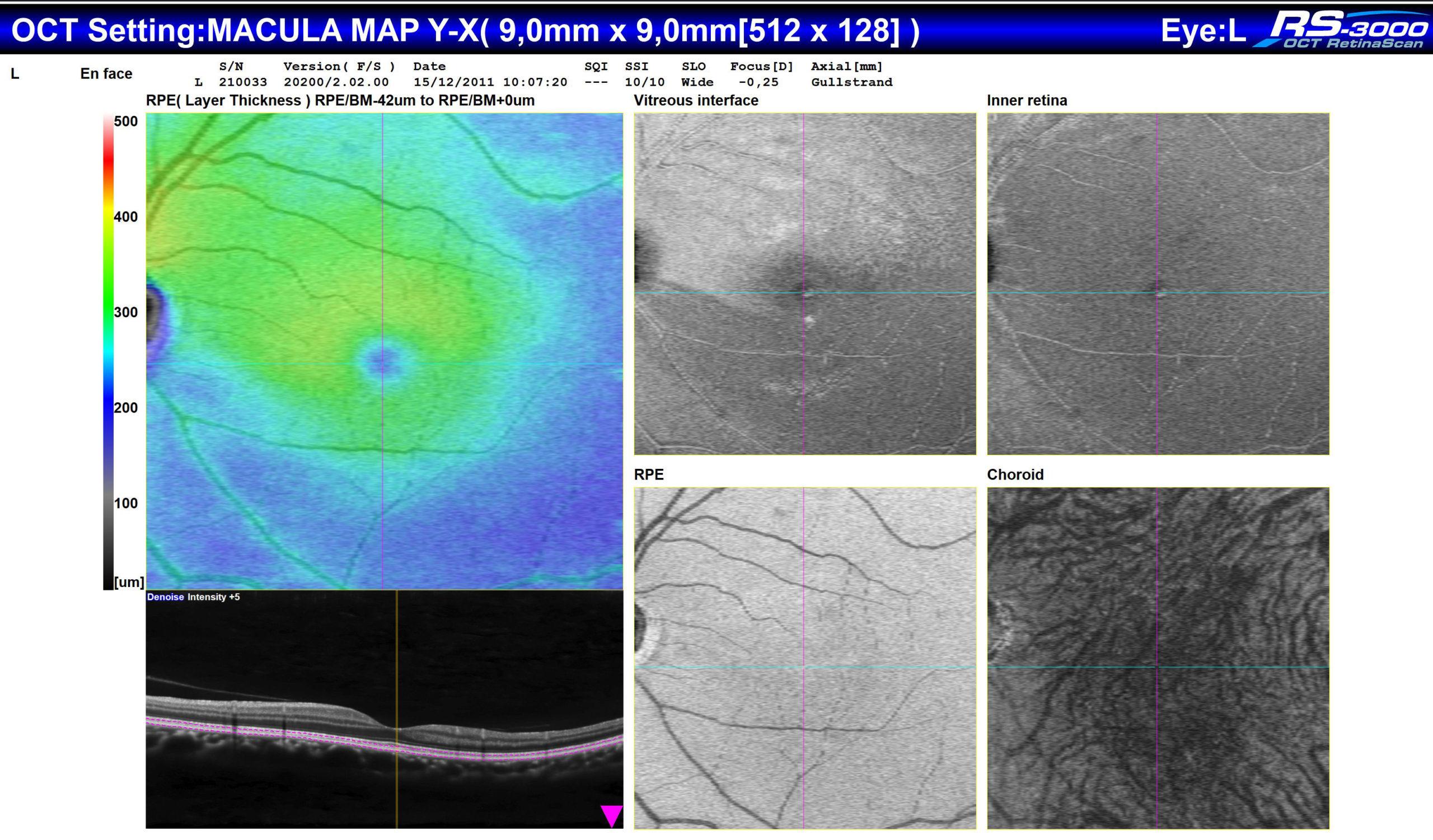
  • Normative database over 9x9 mm on the macula
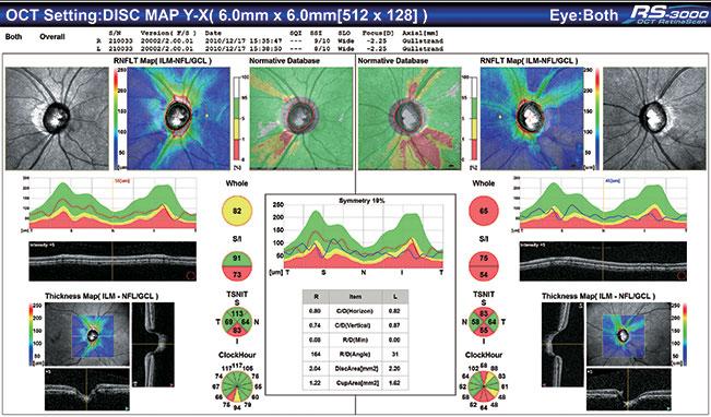
  • RNFL analysis
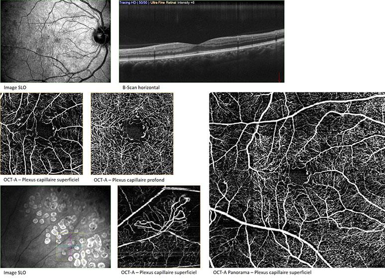
  • Panorama in OCT-Angiography
  • Patient follow-up, up to 50 exams
  • 100% customisable examination reports
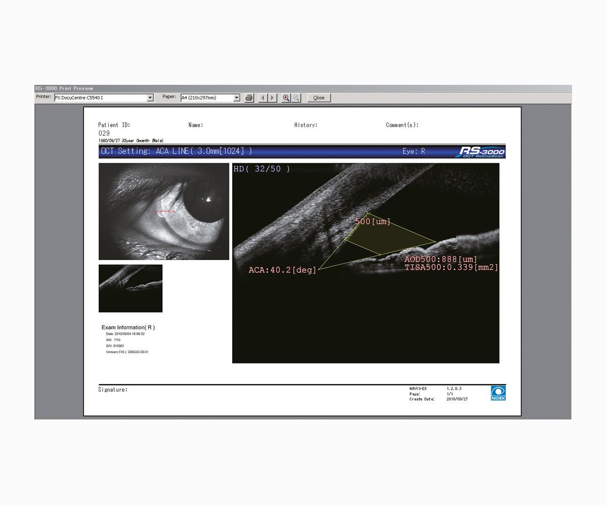
  • Additional lens for OCT sections of the anterior segment

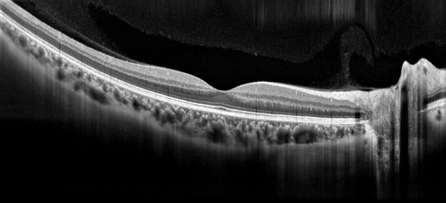
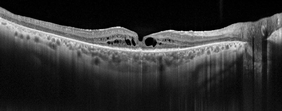
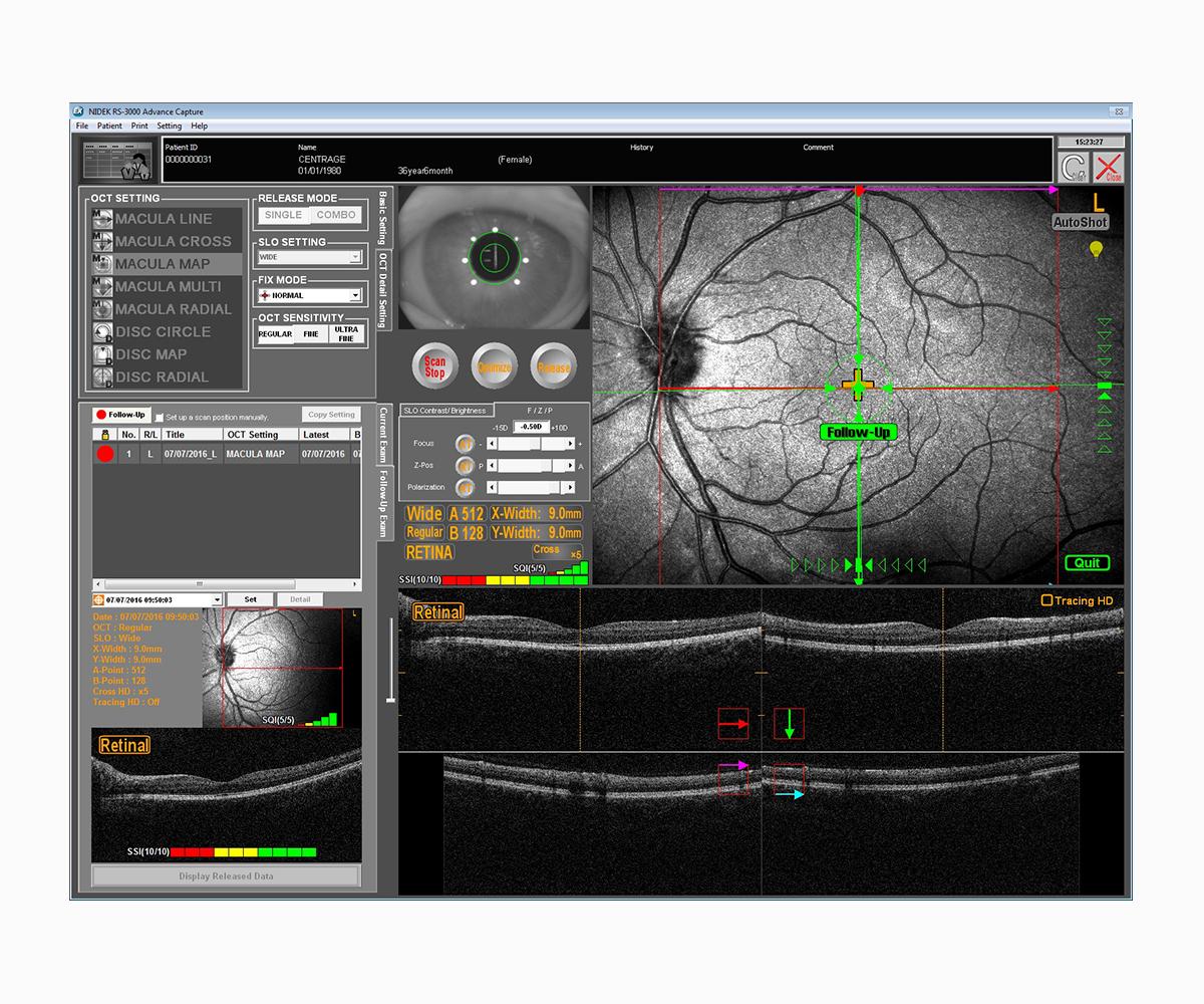
OCT with precise tracking of the retina

The RS-300 Advance 2 comes with the LSO (Laser Scanning Ophthalmoscope) imaging system of the fundus, to track the retina with a high precision, thus ensuring a correct positioning of OCT and OCT-Angiography sections, considering the natural cyclotorsion of the human eye.

This technology makes also possible benefitting from an efficient HD mode, up to 120 scans that, when combined with a maximum sensibility (Ultra-Fine), provides a very accurate image from the vitreous to the choroid, whatever in retina mode or choroid mode (EDI)

Coupe B-scan HD 120 en mode rétine
Coupe B-scan HD 120 en mode choroïde (EDI)
rs 3000 advance 2 tracking disc site web
rs 3000 advance 2 tracking macule site web
Analyse Follow-Up comparative sur Macula Map, complexe des cellules ganglionnaires
Comparative follow-up analysis using Macula Map of the complex of ganglionic cells.
Cartographie d’épaisseur de la cornée
Mapping of cornea thickness
Mesure de l’angle irido-cornéen
Examen de Follow-Up avec reconnaissance de la rétine et repositionnement du scan

Advanced and customisable features

The RS-300 Advance 2 is designed to perform specialised consultations. It benefits from different OCT advanced features, such as the HD mode, the manual positioning of the acquisition scans on the area of interest, the Select mode and the Rescan, to perform an additional exam, or the patient following-up, thanks to Follow-up, available with the acquisition.

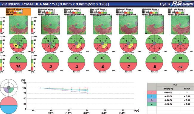
Thanks to the LSO tracking, the RS-300 Advance 2 can perform the following-up exams exactly in the same conditions, using the recognition of the patient’s retina. This ensures a reliable comparative analysis, including on a single section. Over time, up to 50 exams can be compared to monitor the evolution of the pathology and/or to control the efficiency of the cure. By using trend lines and adding manually some events, such the date of the first taking of the treatment for instance, ensuring an efficient and customisable following-up is possible.

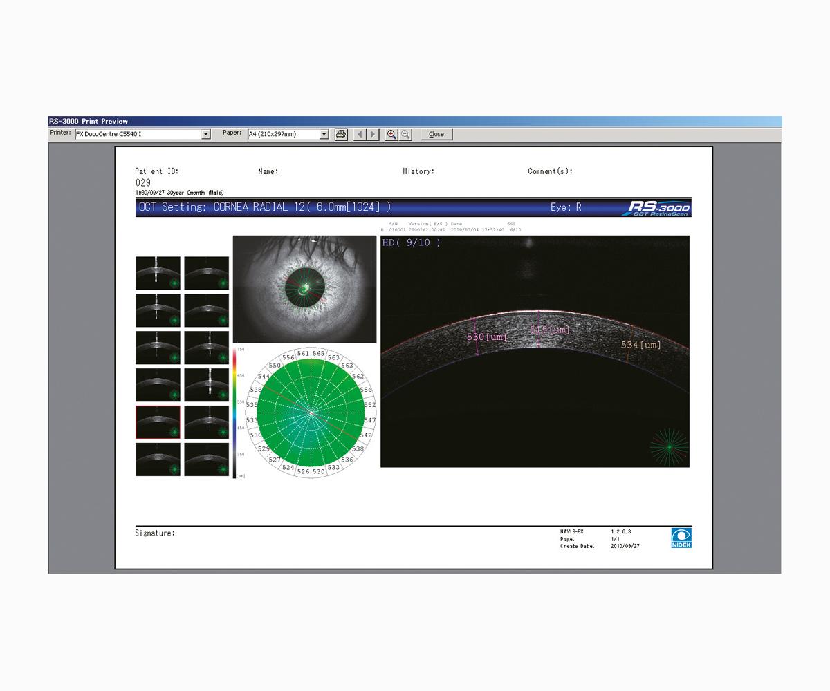
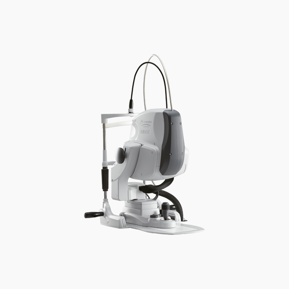
Also, the RS-300 Advance 2 is equipped with an anterior segment adapter to provide sections of the cornea and filtration angles

Result analysis

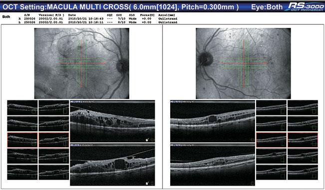
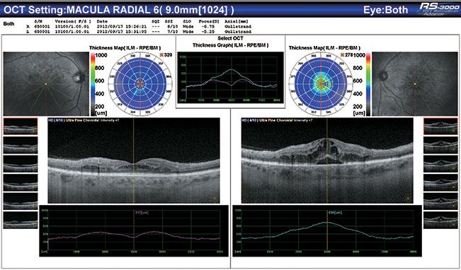
The NAVIS-EX software platform can analyse all the data. This interface receives the viewers dedicated to OCT and OCT-Angiography results and controls the patient database. Different map are displayed: thicknesses, global retina thickness and thickness of the complex of ganglionic cells, these thicknesses being compared with the normative 9x9 mm database, and the thickness of the nervous fibres of the optical nerve, in 6x6 mm. Also displayed are the OCT En-Face and OCT-Angiography representation, all OCT sections and 3D reconstructions.

Epaisseur du complexe des cellules ganglionnaires avec BDN sur 9x9mm (Glaucome)
Analyse du cube 3D ligne par ligne (Macula)
Line-by-line 3D cube analysis (macula)
Cartographie RNFL avec BDN sur 6x6mm (Glaucome)
Analyse OCT En-Face
EN-Face OCT analysis
Résultat du Macula Multi OD/OG
Résultat du Macula Radial OD/OG avec épaisseur de rétine
Analyse OCT-A sur Macula – 7 segmentations
Analyse OCT-A sur Papille – 4 segmentations
Analyse comparative OCT En Face / OCT-A / B-scan sur Macula
Analyse comparative OCT En Face / OCT-A / B-scan sur Papille
Analyse quantitative de densité vasculaire sur Macula
Quantitative analysis of the vascular density on the macula
Analyse quantitative de densité vasculaire sur Papille
Analyse quantitative sur la zone avasculaire centrale

AngioScan: OCT-Angiography module

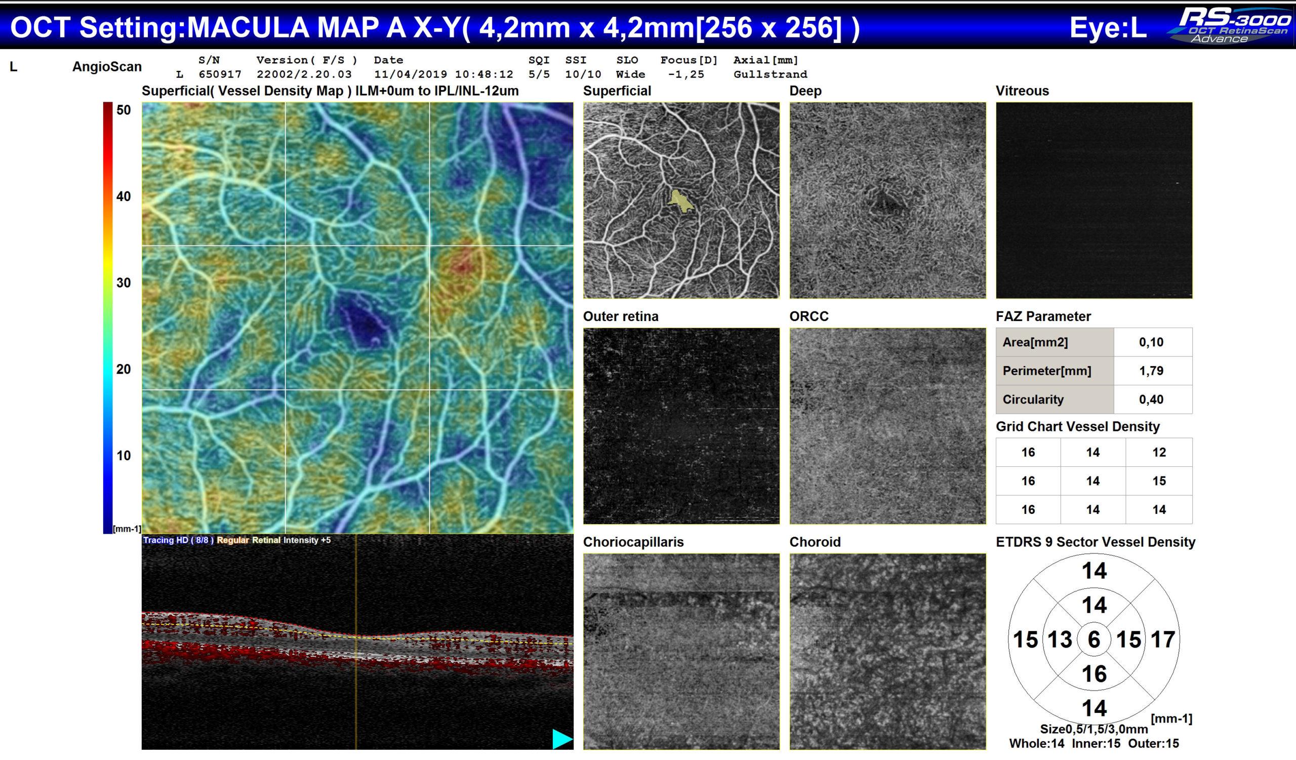
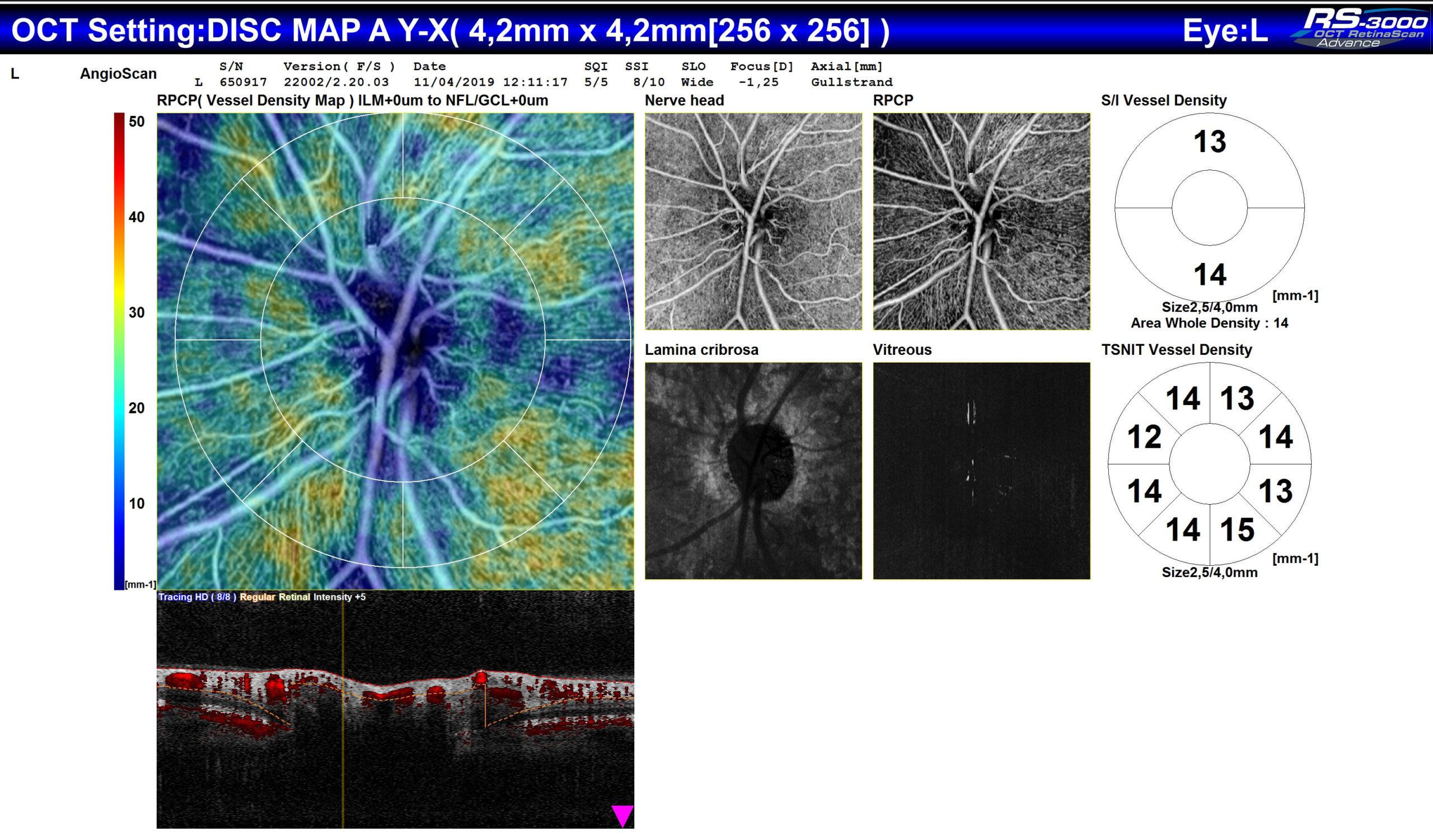
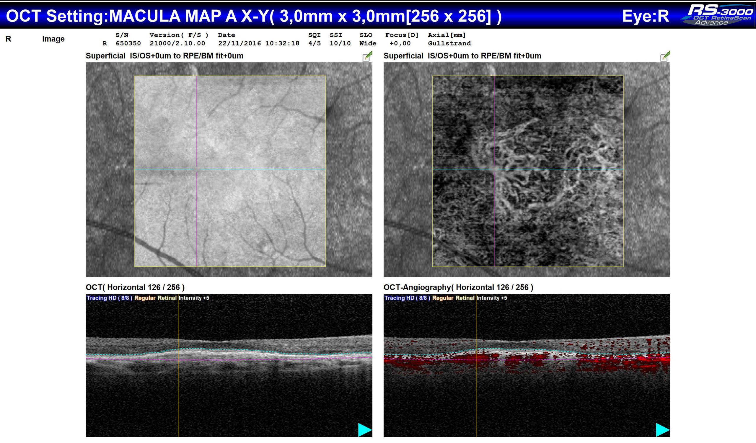
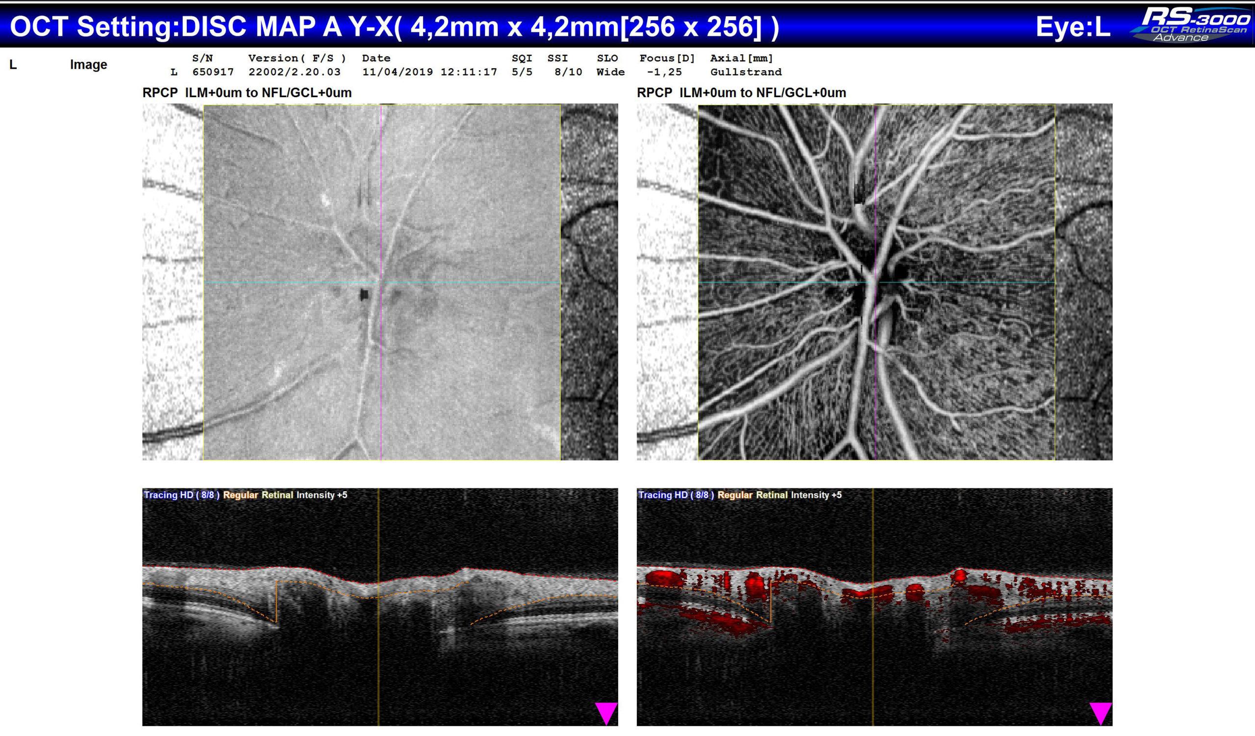
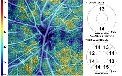
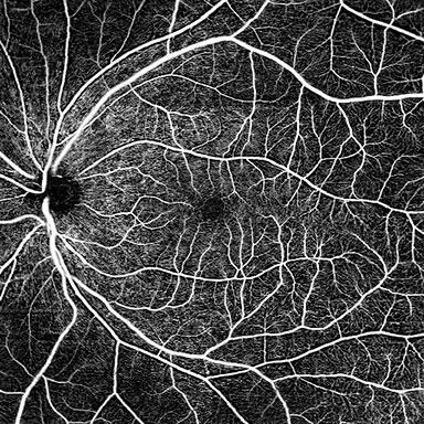
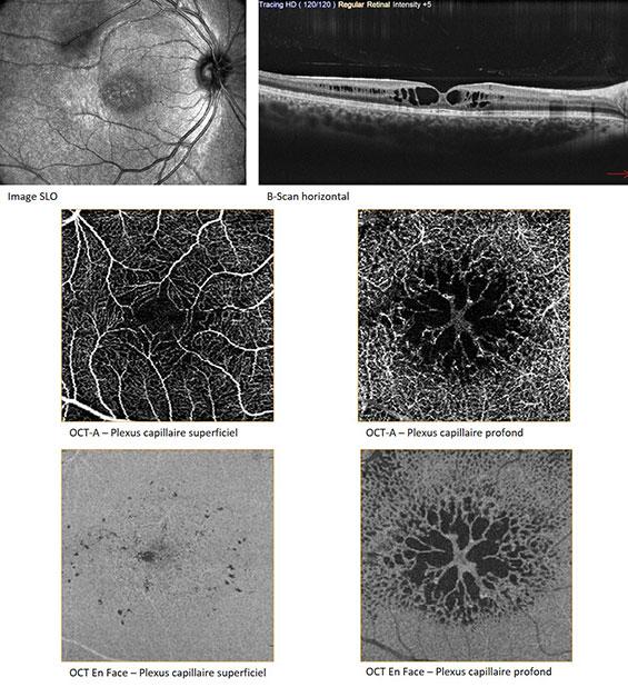
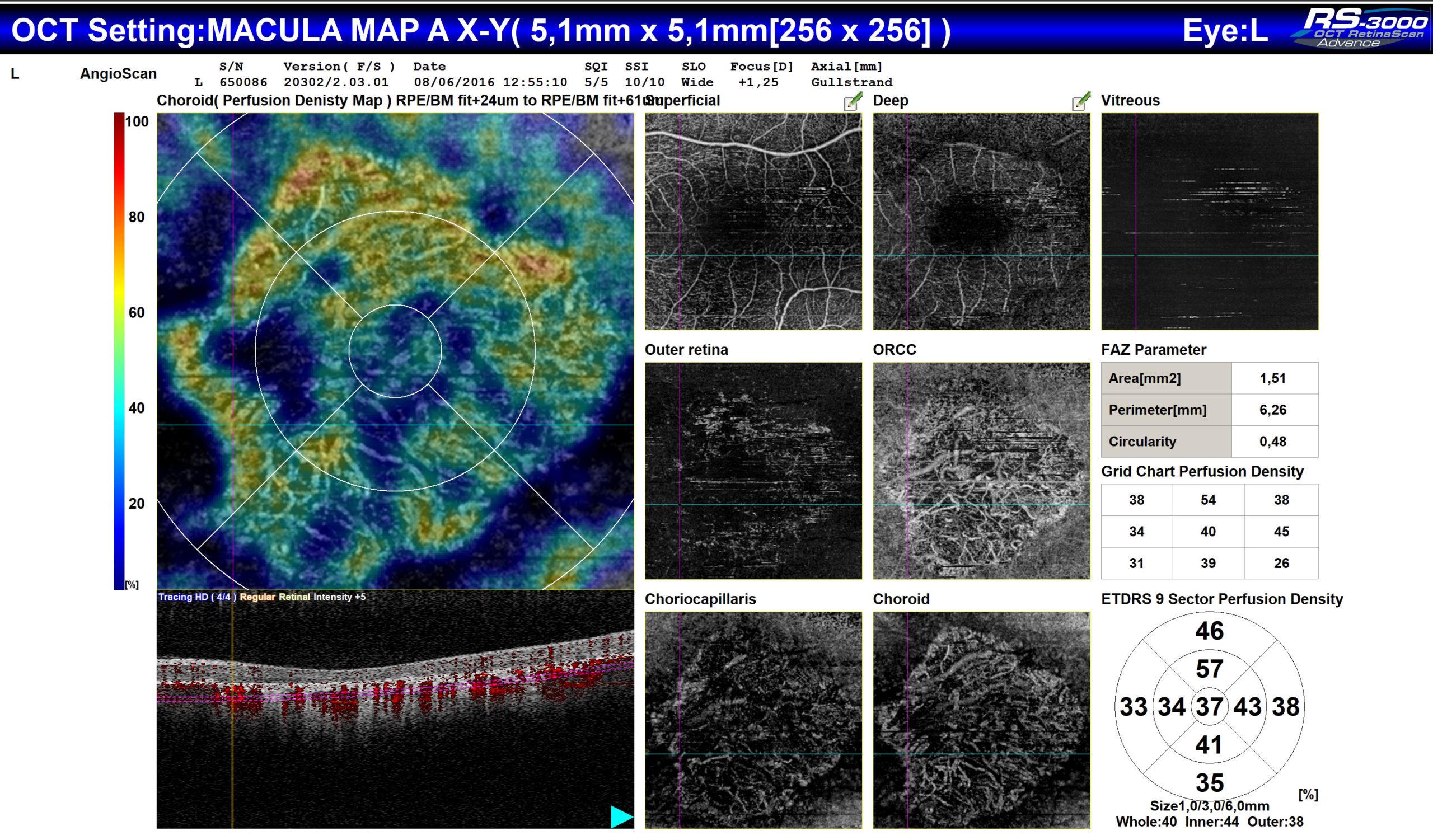
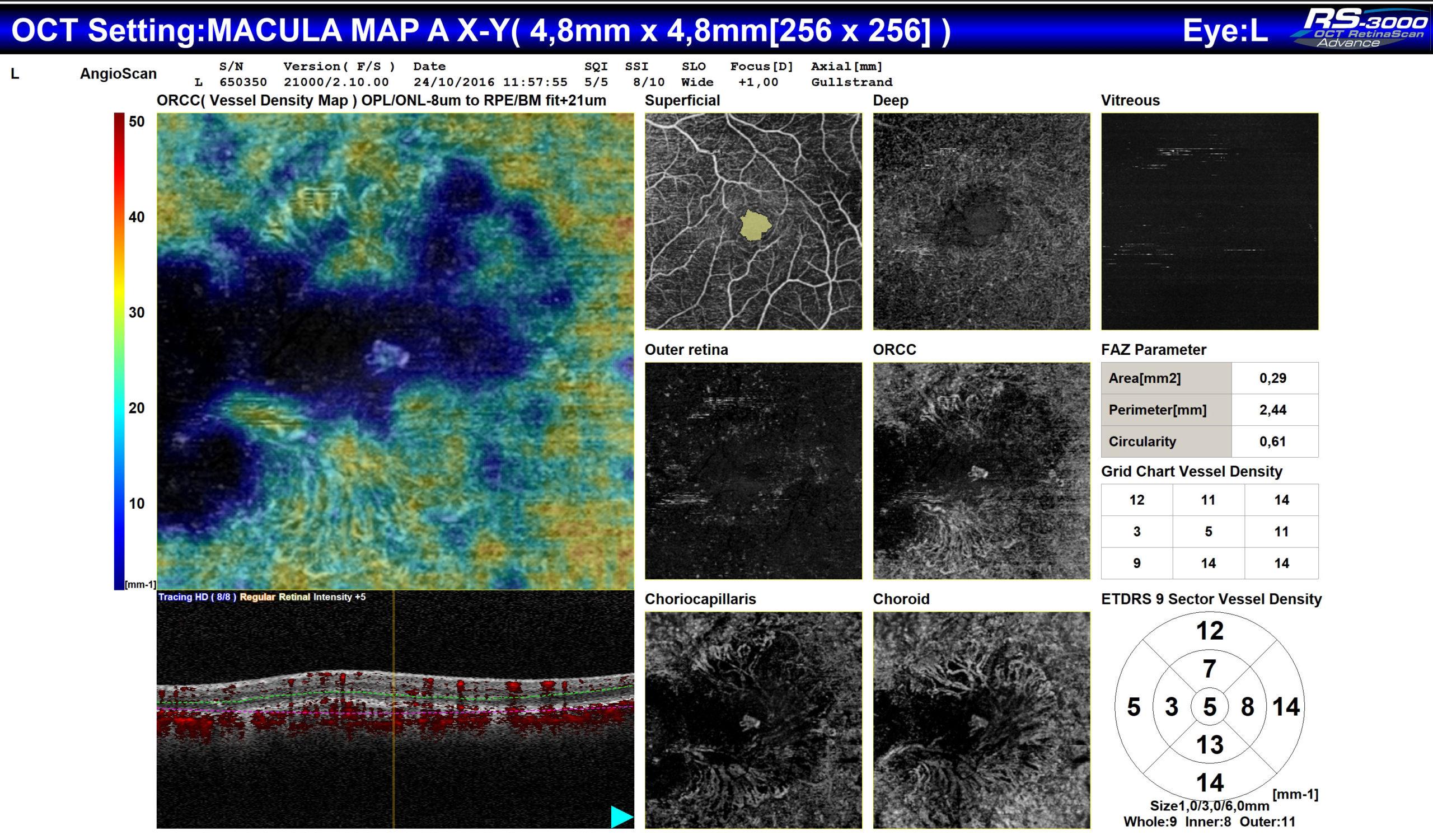
The AngioScan makes possible analysing the vascularisation of the retina and of the choroid. The real time tracking of the retina ensures a correct positioning of the scans, to limit motion artefacts. Images are taken, either of the macula (Macula Map), or of the nervous optical head (Disc Map). The analyses provided are then different. The segmentations are adapted to the captured area, 7 differentiated areas on the macula and 4 areas on the papilla.

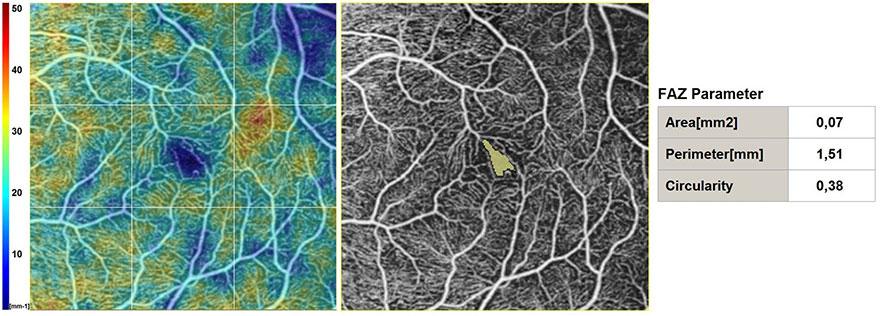
Thanks to the combined OCT En-Face and OCT-Angiography display, whose depth can be adjusted, and to the B-scan section (from a drop-down menu), a direct matching can be made between the structure and the tissue vascularisation.

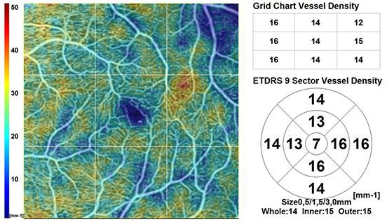
The results of the AngioScan also include quantitative analyses. These additional parameters are used to make a diagnosis. Density and vascular perfusion values are given for each layer, as well as the information related to the central avascular zone (CAZ), which is automatically detected.

Legal mentions

These mentions are conforms to the French regulation and may vary depending on circumstances in each country.

Standard accessories

  • External fixation point
  • Anterior segment adapter with additional front rest.
  • Cleaning air gun
  • Protective cover
  • Protective sheet for chinrest

Consumables

  • Protective sheet for chinrest
  • Good Series RS-3000 cleaning practices

Brochures

  • Sales brochure RS Adv 2
  • Sales brochure AngioScan
  • Livret OCT-Angiographie


Livrets de cas cliniques

  • Clinical case book Dr.
  • Clinical case book Dr.
  • Clinical case book Prof. Lida


OCT Denoising

  • A New Solution for Overcoming OCT Noise – Rodrigo Abreu Gonzales, MD, PHD, FEBO

OCT-A

  • Correlation of OCT Angiography and Microperimetry in Wet AMD – anish Nagpal, MS, DO, FRCS(Edin); Jayesh Khandelwal, MS; Rakesh Juneja, MS; and Navneet Mehrotra, MBBS, DNB (Ophthalmology), FRF
  • OCT Angiography in Retinal and Choroidal Diseases – Manish Nagpal, MS, DO, FRCS(Edin), and Shishir Shekhar Singh, MS
  • The Role of OCT Angiography in Assessing Retinal and Choroidal Vasculature – DANIELA BACHERINI, MD, PHD
  • Panoramic Imaging With OCTA – Manish Nagpal, MS, DO, FRCS(Edin), and Shishir Shekhar Singh, MS
  • Peripapillary Vascular Changes in Radiation Optic Neuropathy : An Optical Coherence Tomography Angiography Grading – Raffaele Parrozzani, Luisa Frizziero, Davide Londei, Sara Trainiti, Rocco Luigi Modugno, Francesca Leonardi, Serena Pulze, Giacomo Miglionico, Elisabetta Pilotto, Edoardo Midena
  • Morphological Changes And Quantitative Analysis Of Macular Retinal Microvasculature By Optical Coherence Tomography Angiography In Hypertensive Retinopathy – Yinhe Liu, Jushuang Li, Jian Pan, Yonghua Wang, Guangyun Mao & Zipei Jiang


OCT-A et Micropérimétrie

  • Structural and functional retinal changes in patients with type 2 diabetes without diabetic retinopathy – Qiannan Chai, Yimin Yao, Congrong Guo, Hong Lu, and Jingxue Ma
    https://www.ncbi.nlm.nih.gov/pmc/articles/PMC9258434/
  • Multimodal Structure and Function Analysis – Nadia K. Waheed, MD, MPH
THE OFFER OF NIDEK PRODUCTS

Produits complémentaires du RS-3000 Advance 2

Loading...

MP-3

FIND MORE

NAVIS-EX Software

FIND MORE
PRODUCTS YOU MAY ALSO BE INTERESTED IN

In the same range

Loading...

RS-330

FIND MORE

Mirante (SLO/OCT)

FIND MORE

Do you require more information?

You have a project? You want a quotation? You have questions about our products?
Feel free to ask your technical sales representative.

Contact us

The art of Eye Care

Reliability and
safety

NIDEK develops its top-of-the-range products to improve visual health through an approach based on strict criteria: safety, reliability, durability, continuous quality controls and certifications.

Technologies and
innovations

NIDEK meets technical challenges by keeping constantly informed of the innovations of eye imaging systems, using the expertise of professionals and the progresses of research.

Services and
guarantees

NIDEK commits itself to providing services to its customers, from the installation of an activity to the authorised training of teams, and to offering long-time measurable guarantees.
FIND MORE

Contact NIDEK

Contact us

Spécialiste des équipements et technologies au service de la santé visuelle, NIDEK SA accompagne et équipe les ophtalmologistes, orthoptistes, professionnels de l’optique et les établissements de santé.

Facebook Linkedin Youtube

KEY PRODUCTS

  • Mirante
  • TS 610
  • RT-Connect
  • Synetic

Services

  • Design office
  • Installation & Implementation
  • Trainings
  • ASS and repairing

Solutions

  • Optics
  • Ophthalmology
  • Orthoptics
  • Health care establishment

About us

  • Expertise and know-how
  • Our key dates
  • Certifications
  • Nidek in France and worldwide
  • Recruitment

© 2021 • NIDEK SA – All rights reserved

  • General Terms and Conditions
  • Protection des données personnelles
  • Legal notices
Gérer le consentement aux cookies
Nous utilisons des cookies pour optimiser notre site web et notre service.
Fonctionnel Always active
Le stockage ou l’accès technique est strictement nécessaire dans la finalité d’intérêt légitime de permettre l’utilisation d’un service spécifique explicitement demandé par l’abonné ou l’utilisateur, ou dans le seul but d’effectuer la transmission d’une communication sur un réseau de communications électroniques.
Préférences
Le stockage ou l’accès technique est nécessaire dans la finalité d’intérêt légitime de stocker des préférences qui ne sont pas demandées par l’abonné ou l’utilisateur.
Statistiques
Le stockage ou l’accès technique qui est utilisé exclusivement à des fins statistiques. Le stockage ou l’accès technique qui est utilisé exclusivement dans des finalités statistiques anonymes. En l’absence d’une assignation à comparaître, d’une conformité volontaire de la part de votre fournisseur d’accès à internet ou d’enregistrements supplémentaires provenant d’une tierce partie, les informations stockées ou extraites à cette seule fin ne peuvent généralement pas être utilisées pour vous identifier.
Marketing
Le stockage ou l’accès technique est nécessaire pour créer des profils d’utilisateurs afin d’envoyer des publicités, ou pour suivre l’utilisateur sur un site web ou sur plusieurs sites web ayant des finalités marketing similaires.
Manage options Manage services Manage {vendor_count} vendors Read more about these purposes
Préférences
{title} {title} {title}

Galerie

OCT-A Panorama en 12×12 mm
Cas de Glaucome – Analyses d’épaisseur et de densités vasculaires sur macula (cellules ganglionnaires) et sur la tête du nerf optique (RNFL)
Analyses OCT et OCT-Angiographie (région maculaire et périphérique) dans un cas de rétinopathie diabétique
Analyses OCT et OCT-Angiographie dans un cas de rétinoschisis lié à l’X
Rapport d’examen OCT-Angiographie dans un cas de dégénérescence maculaire liée à l’âge atrophique
Rapport d’examen OCT-Angiographie dans un cas de rétinopathie diabétique proliférative avec néovaisseau papillaire
Rapport d’examen OCT-Angiographie dans le cas d’un néovaisseau de type 1
Rapport d’examen OCT-Angiographie dans un cas d’un néovaisseau de type 2

Call me back

A problem was detected in the following Form. Submitting it could result in errors. Please contact the site administrator.

Télécharger votre brochure

Télécharger votre brochure

Télécharger votre brochure

Télécharger votre brochure

Télécharger votre brochure

Télécharger votre brochure

Télécharger votre brochure

Télécharger votre brochure

Télécharger votre brochure

Télécharger votre brochure

Télécharger votre brochure

Télécharger votre brochure

Télécharger votre brochure

Télécharger votre brochure

Télécharger votre brochure

Télécharger votre brochure

Télécharger votre brochure

Télécharger votre brochure

Télécharger votre brochure

Télécharger votre brochure

Télécharger votre brochure

Télécharger votre brochure

Télécharger votre brochure

Télécharger votre brochure

Ce site est réservé aux professionnels de santé

Sortir
Je suis professionnel de santé