• 01 49 80 97 97
  • Contact us
  • Boutique NIDEK

Efficient After Sales Service

At your disposal whatever the situation

Made in France

Our consultation units

Our experts at your disposal

60 experts over the territory

  • Lasers
        • Lasers YAG and SLT
          • YC-200 S Plus (YAG/SLT)
          • YC-200 (YAG)
        • Photocoagulator lasers
          • YLC-500 / YLC 500 VIXI
          • GYC-500 / GYC 500 VIXI
  • Retina diagnosis
        • Slit lamps
          • SL-9900/SL-9900 Zoom
          • SL-9800
          • SL-250
        • Multimodal platforms
          • Mirante (SLO/OCT)
          • Mirante SLO (SLO)
        • Fundus cameras
          • AFC-330
          • Retina Scan Duo 2
          • Mirante SLO (SLO)
          • Mirante (SLO/OCT)
        • Microperimeters
          • MP-3
          • MP-3S
        • Angiographs
          • Mirante (SLO/OCT)
          • Mirante SLO (SLO)
        • Software
          • NAVIS-EX Software
        • OCT
          • RS-1
          • Retina Scan Duo 2
          • Mirante (SLO/OCT)
        • Ultrasound scanners
          • US-4000/4000-P (Echo A+B)
          • US-500 / 500-P (Echo A)
  • Anterior segment
        • Slit lamps
          • SL-9900/SL-9900 Zoom
          • SL-9800
          • SL-250
        • Aberrometers - Topographs
          • OPD-Scan III
        • Optical biometers
          • AL-Scan
          • AL-Scan M
        • Tonometers
          • TONOREF III
          • NT-1e / NT-1 / NT-1p
        • Specular microscope
          • CEM-530
        • Goniolenses
          • GS-1
  • Refraction
        • Pupillometers
          • PM-700
        • Afficheurs de tests
          • CP-9
          • SC-1600 Pola/SC-1600
        • Itinerant optician case
          • Itinerant optician case
        • Lensometers
          • LM 1800 P – PD
          • LM7 – 7P
        • Auto refractometers
          • AR-1/ARK-1
          • AR-1a/ARK-1a
          • AR-1s/ARK-1s
          • AR-F/ARK-F
          • HandyRef/HandyRef-K
          • TONOREF III
        • Refractors
          • RT-3100
          • TS-310
          • RT-6100
          • TS-610
          • Logiciel RT-Connect
        • Système de réfraction assisté
          • FARS (Fully Assisted Refraction System)
  • Consultation units
        • "Excellence" units
          • Synetic
          • OT-6400
        • "Performance" units
          • Affinity
          • OT-4200
        • "Compact" units
          • ONE-i
          • Lypop
          • COS-6100
        • Lifting tables and examination swivel chair
          • Consultation armchairs and stools
          • Tables à élévation Up
  • Optical workshop
        • Automatic edges
          • ME-1500
          • LEXCE Trend 8D
          • LEXCE Trend D
          • LEXCE Trend DBT
          • SE9090 Supra
        • Lensometers
          • LM 1800 P – PD
          • LM7 – 7P
        • Centering-blocking devices
          • ICE-1500
          • ICE- 1
        • Shape plotters
          • LT-980
          • LETR-3 Traceur LEXCE Trend
        • Green products
          • LFU-220
          • LED-200 Plus
        • Colonne de centrage : Invision Fit AI
  • NIDEK
        • Our services
          • Design office
          • Installation & Implementation
          • Trainings
          • ASS and repairing
          • Environement
          • Showroom NIDEK
        • Our solutions
          • Optics
          • Ophthalmology
          • Orthoptics
          • Health care establishment
        • About us
          • Expertise and know-how
          • Certifications
          • NIDEK in France
          • NIDEK worldwide
          • Recruitment
        • News
          • Blog
          • Évènements
  • Our shop
  • fr_FR
  • en_GB
PageProduit_RS330_VisuelProduit_Intro

Homepage > OCT > RS-330

RS-330

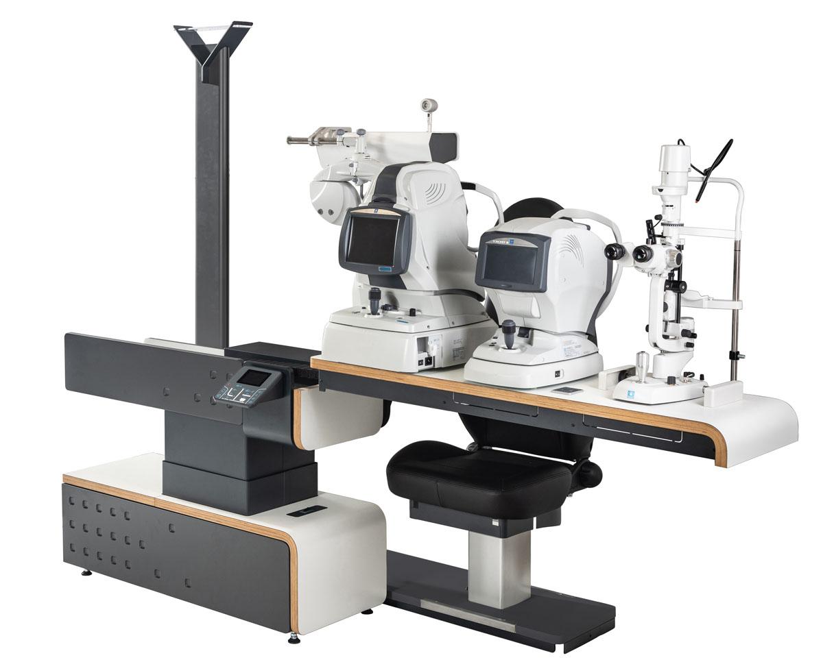
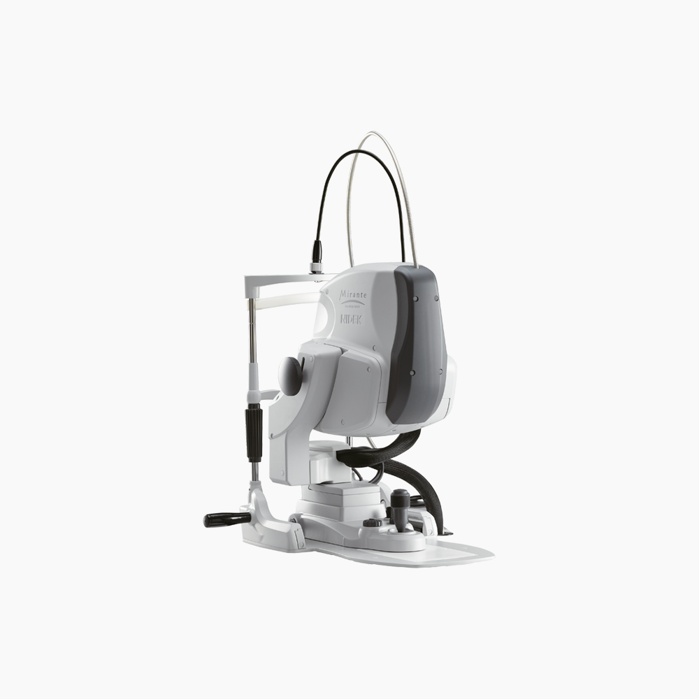
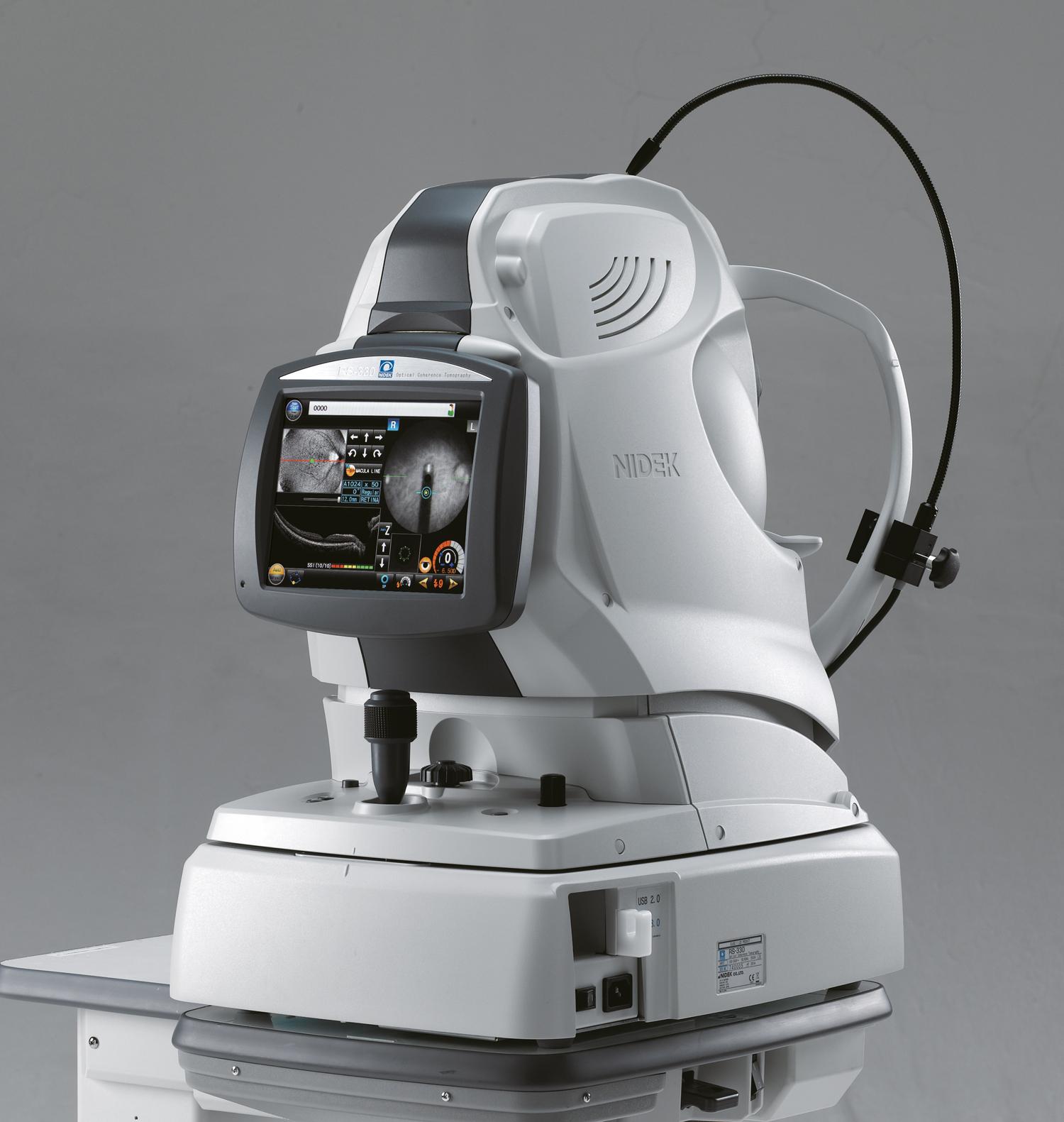
The RS-330 is a combined OCT used to analyse sections of the retina, to make pictures and to examine the fundus auto fluorescence. Thanks to an additional lens, it also makes possible analysing sections of the anterior segment and making pictures of it. Therefore it is a powerful detection tool able to detect early the eye lesions. 

  • More details
  • Get to the gallery
Call me back
Download the brochure
  • Summary
  • Description
  • Accessories
  • Cleaning
  • Documentation
  • OCT, non-mydriatic fundus camera, and auto fluorescence combined system
  • 53,000 A-scans/s speed
  • Built-in 12 Mega pixel 45° camera
  • Colour photo of the anterior segment
  • Panoramic reconstruction with 7 ETDRS dials
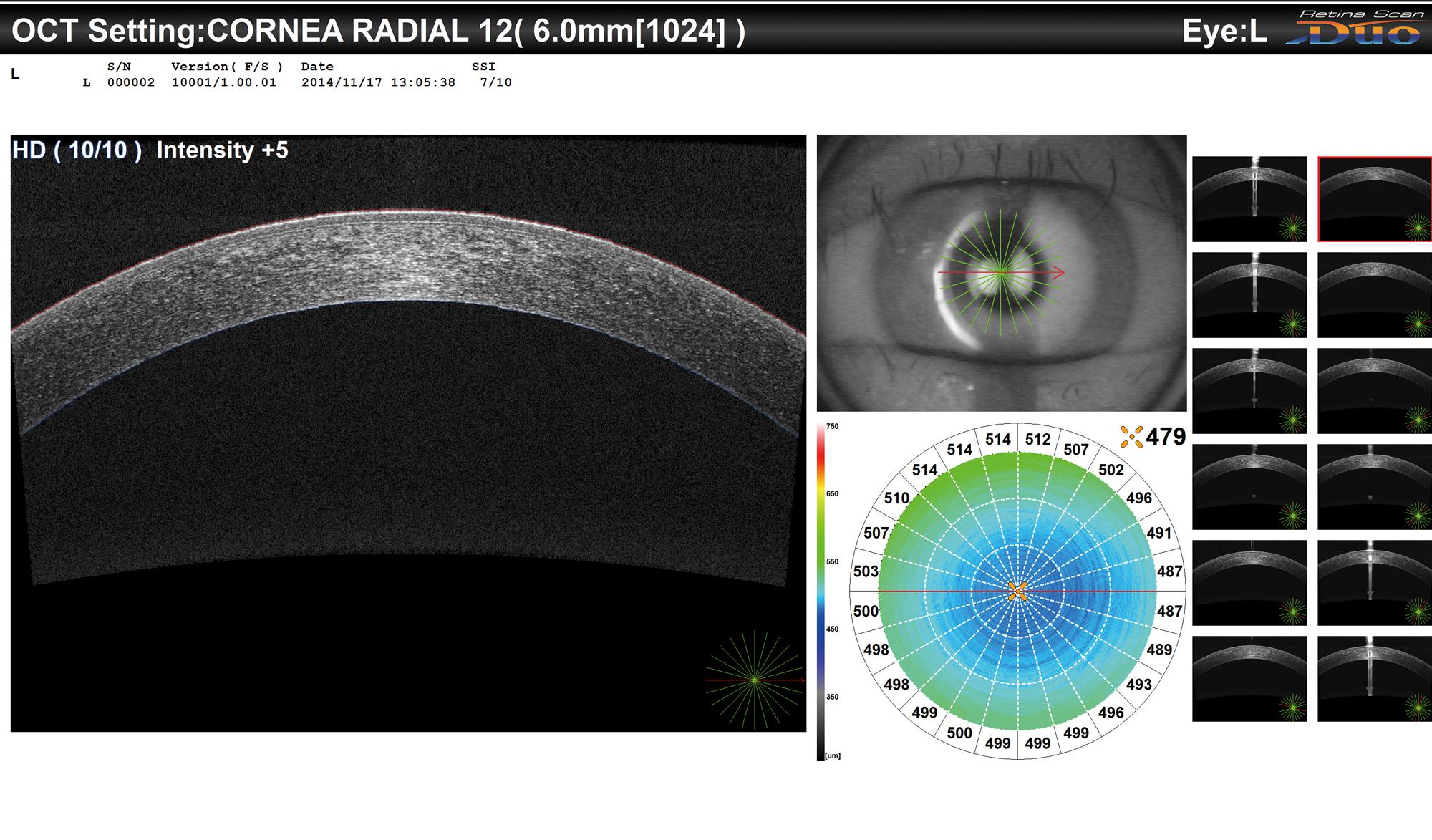
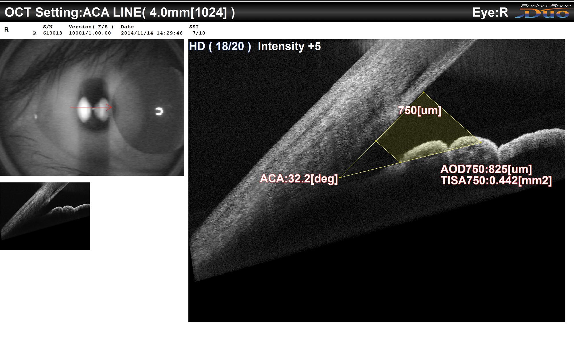
  • Additional lens to perform OCT sections od the anterior segment
  • 3D alignment, automatic focusing and triggering to make the device very easy to use
  • Initial dioptrical compensation from -12D to +10D
  • COMBO mode to delegate the tasks
  • Modulation of the signal sensibility for opaque media

Detection tool to delegate the tasks

The RS-330 combines the OCT, the RNM and the auto fluorescence. It is equipped with a 3D tracking system of the eye and with different automatisms to make the collection of images easier.

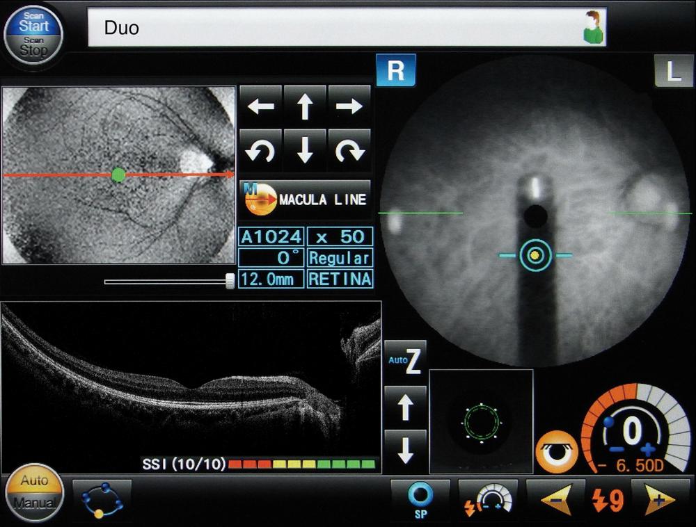
To be user-friendly, the device tracks the patient’s eye in real time, performs the alignment with the pupil, the focus on the retina and triggers the acquisition (OCT + photo). Combined with the COMBO’s, the RS-330 performs series of customisable examinations, programmed depending on the needs (ex: Macula Map + Disc Map for glaucoma). It is perfectly adapted to a routine use, to make the detection and standardise the examinations. The signal sensibility can be changed (Regular, Fine or Ultra-Fine mode). This modifiable parameter strengthens the intensity of the signal in opaque media.

Depending on the configuration of the consultation, you can install the device on a NIDEK dedicated lifting table (depending on the model) for a daily use.

Fenêtre d’acquisition
Exemple d’installation sur la Synetic NIDEK (unité à translation)
PageProduit_RS330_Description_Visuel3_4_5
Cartographie d’épaisseur de la cornée
Mapping of cornea thickness
Mesure de l’angle irido-cornéen

Variety of measurements

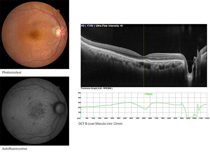
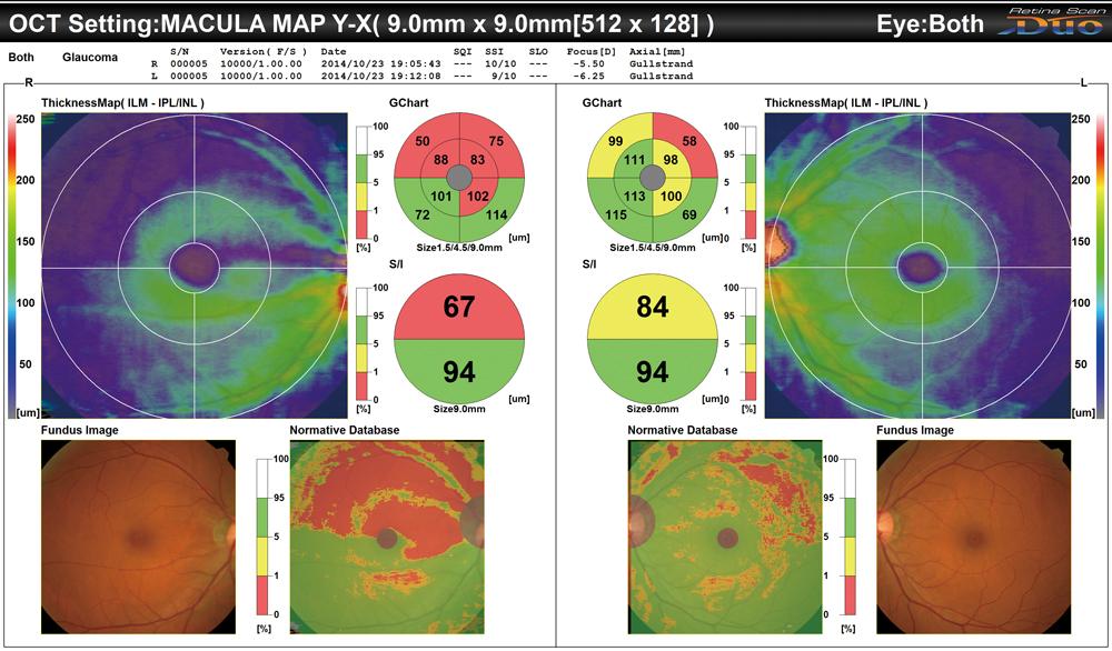
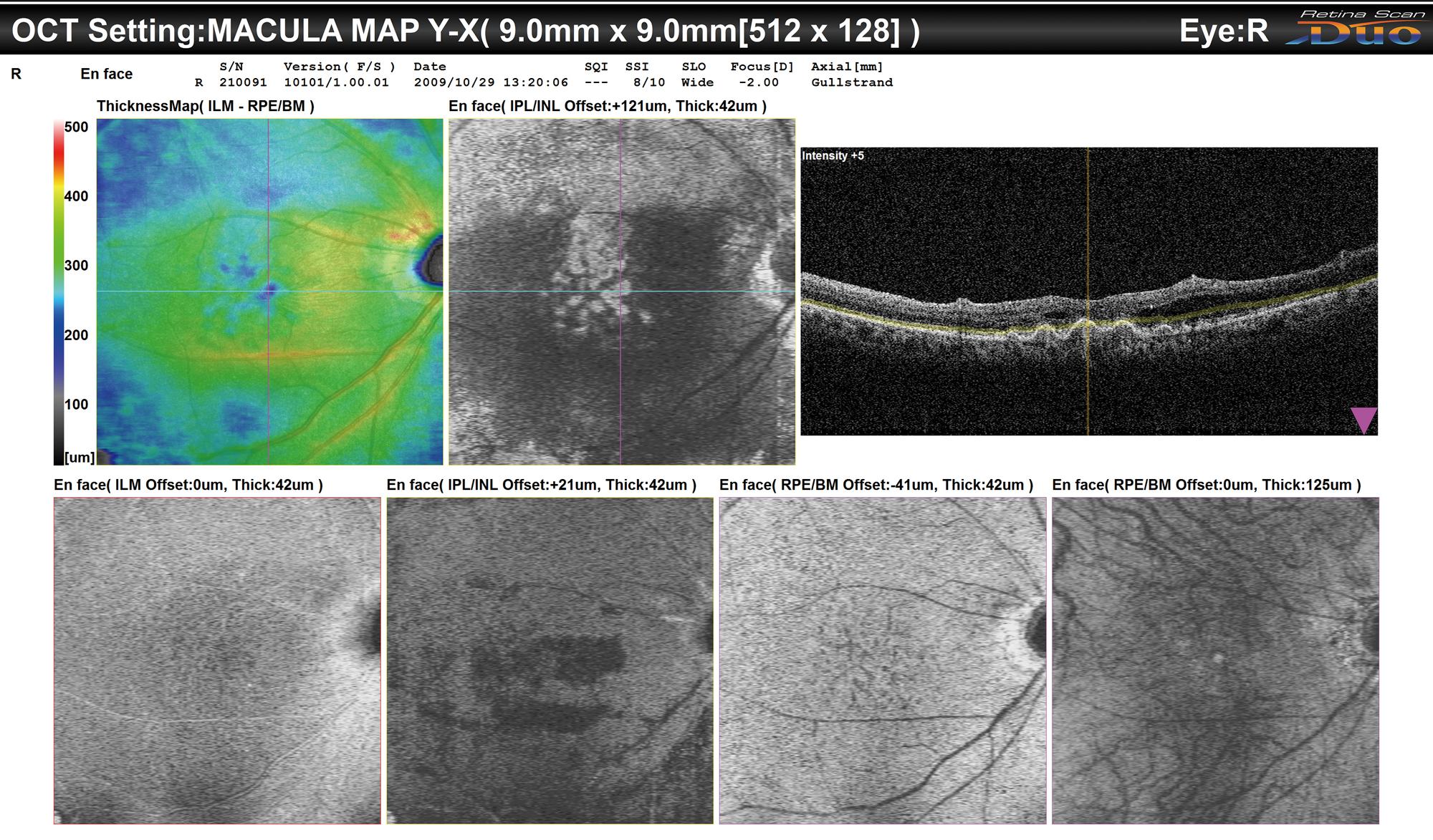
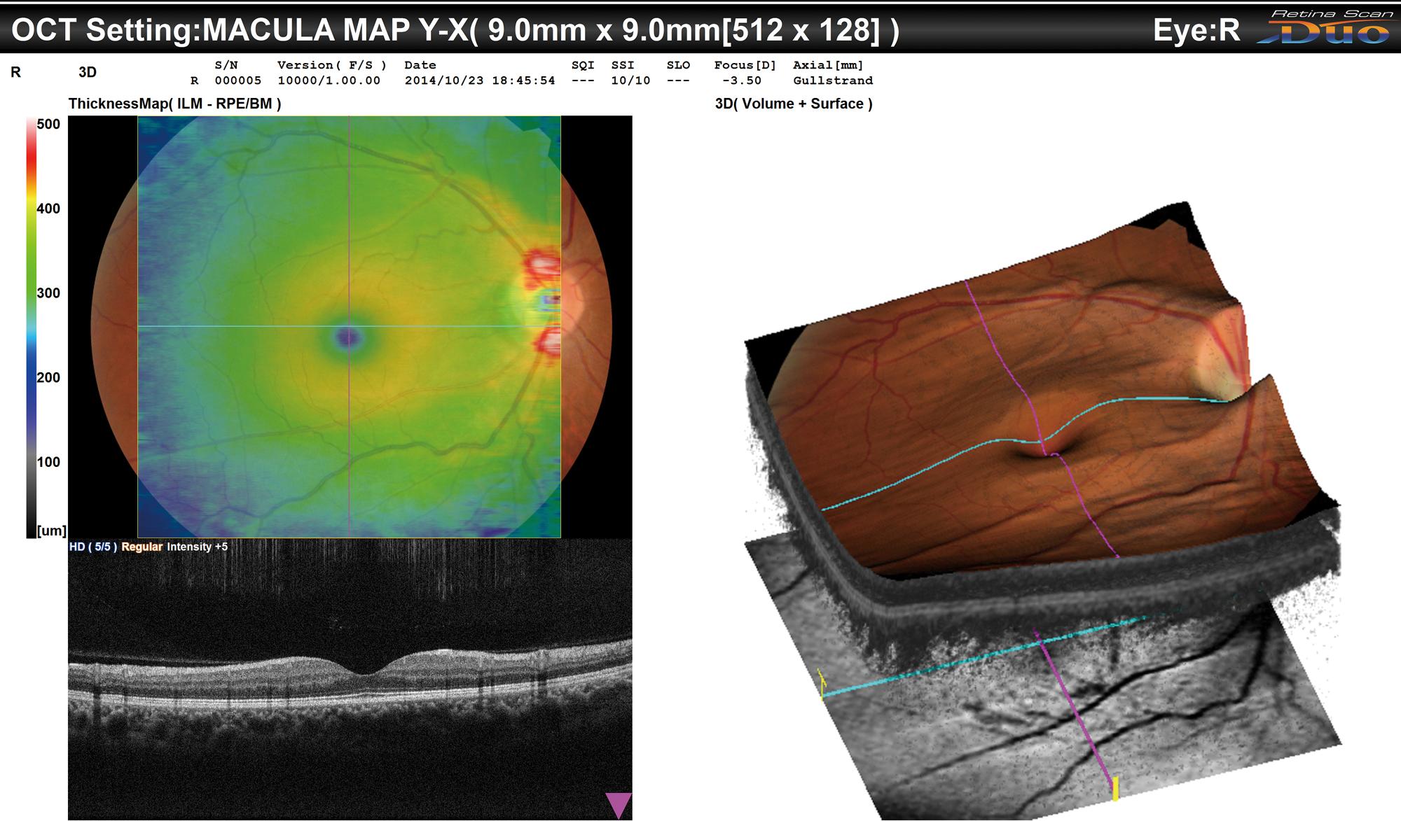
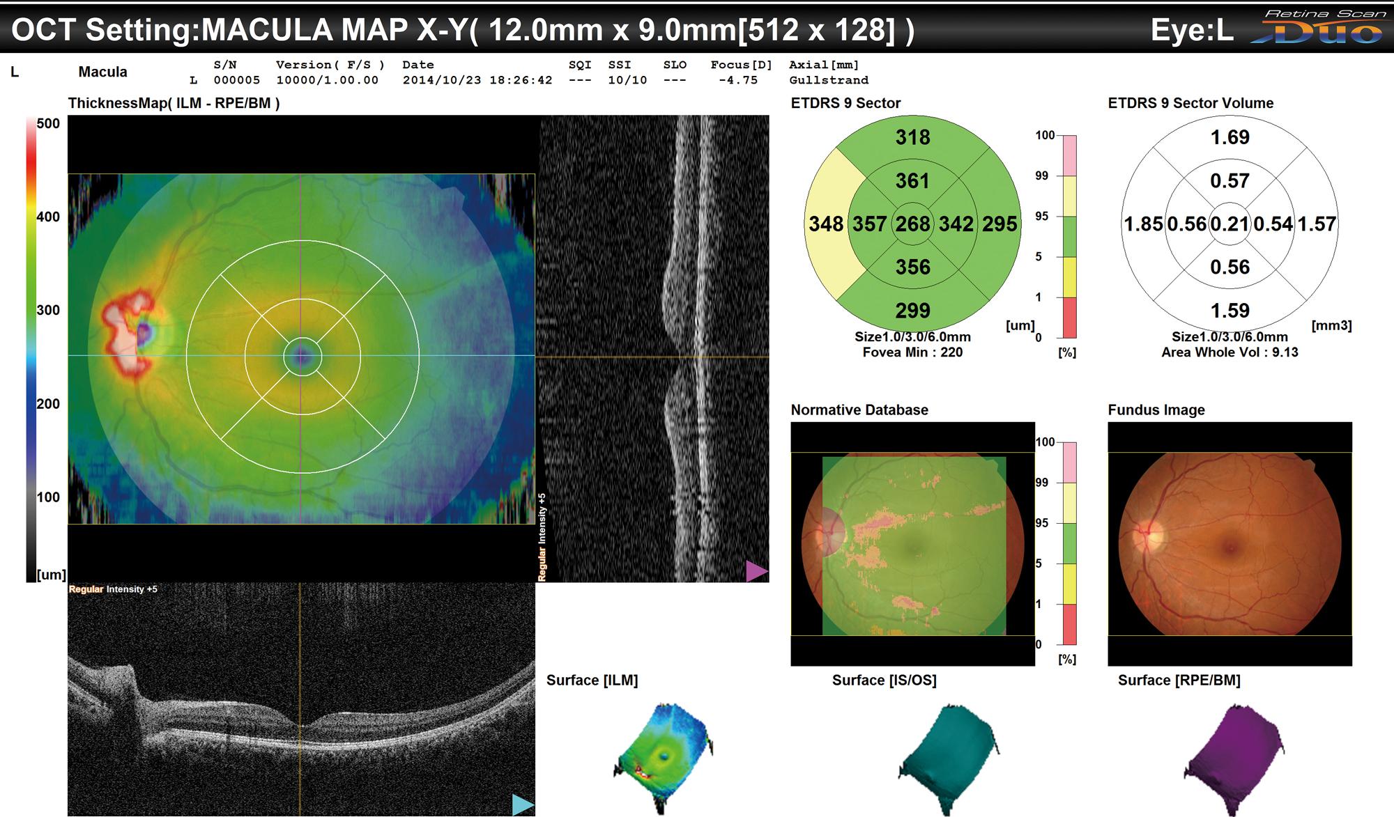
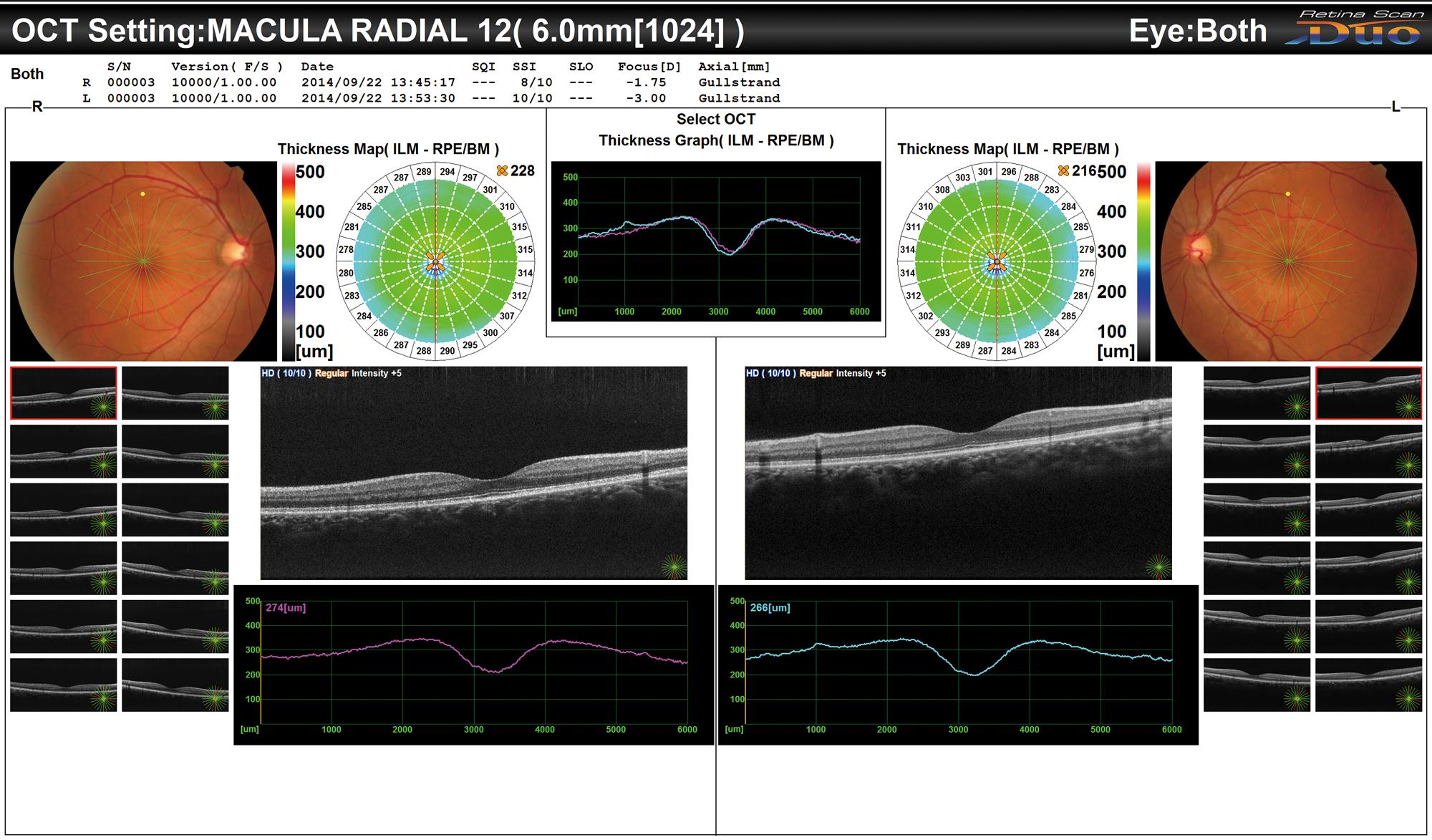
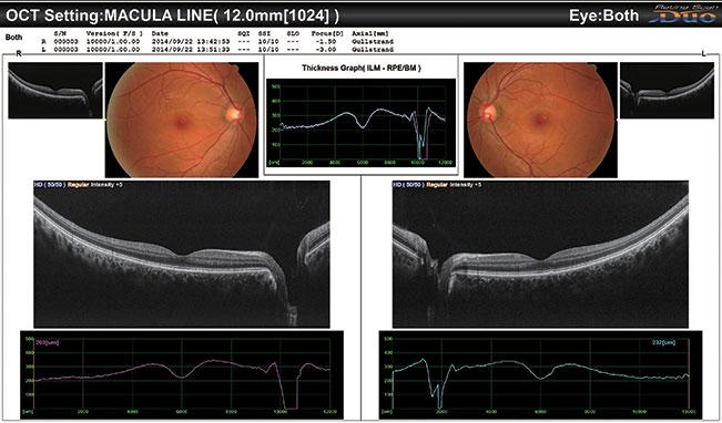
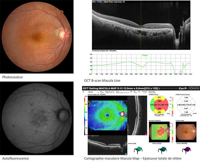
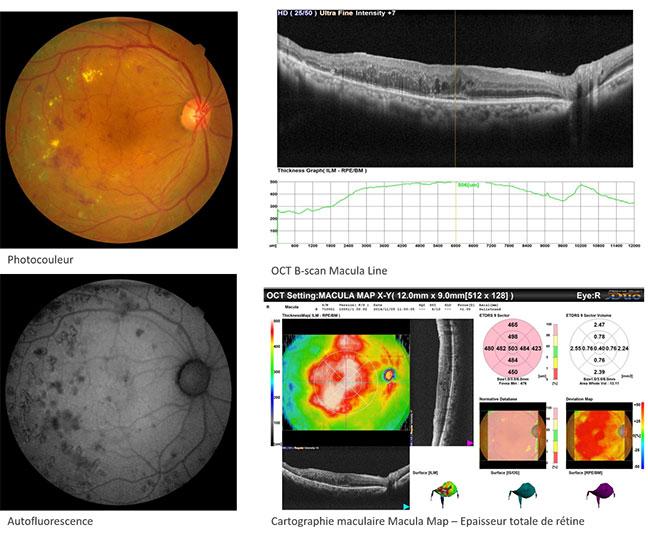
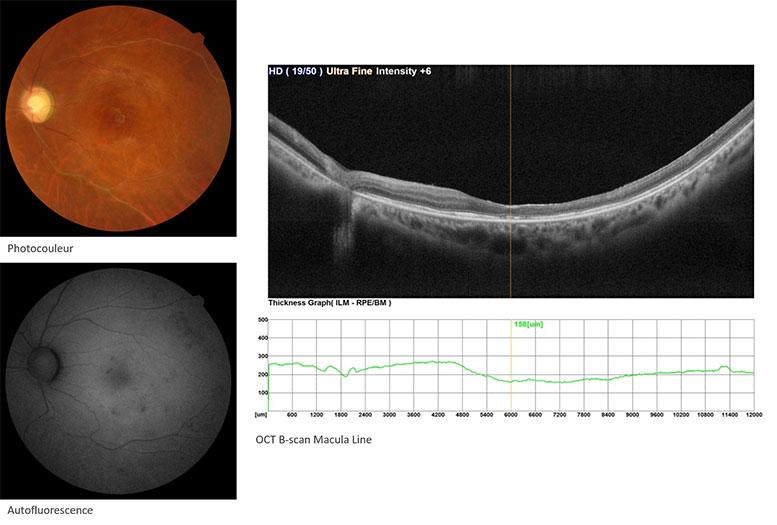
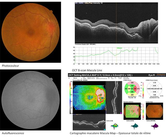
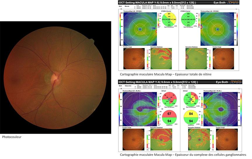
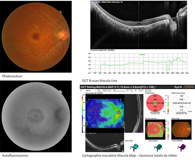
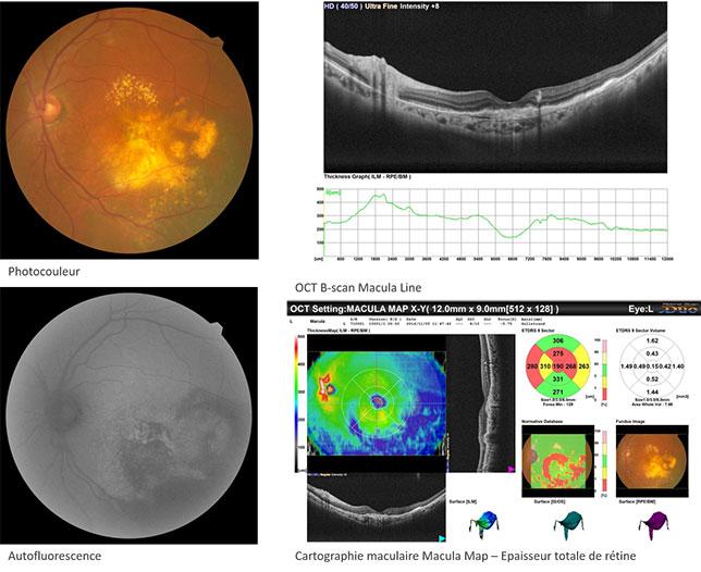
The acquisition fields, in fundus photography and auto fluorescence, reach 12x9 mm, in OCT, and 45° in fundus photography and auto fluorescence. They provide a multimodal analysis of the posterior pole and enable an early detection of some macula diseases, thanks to the green wavelength auto fluorescence, because the macula pigment less absorbs it.

To perform a further expertise, different other imaging modalities are available: different OCT sections, image summation up to 50 sections (HD mode), EDI mode to view the choroid or colour photo and anterior segment OCT modes.

Adapted analysis tools

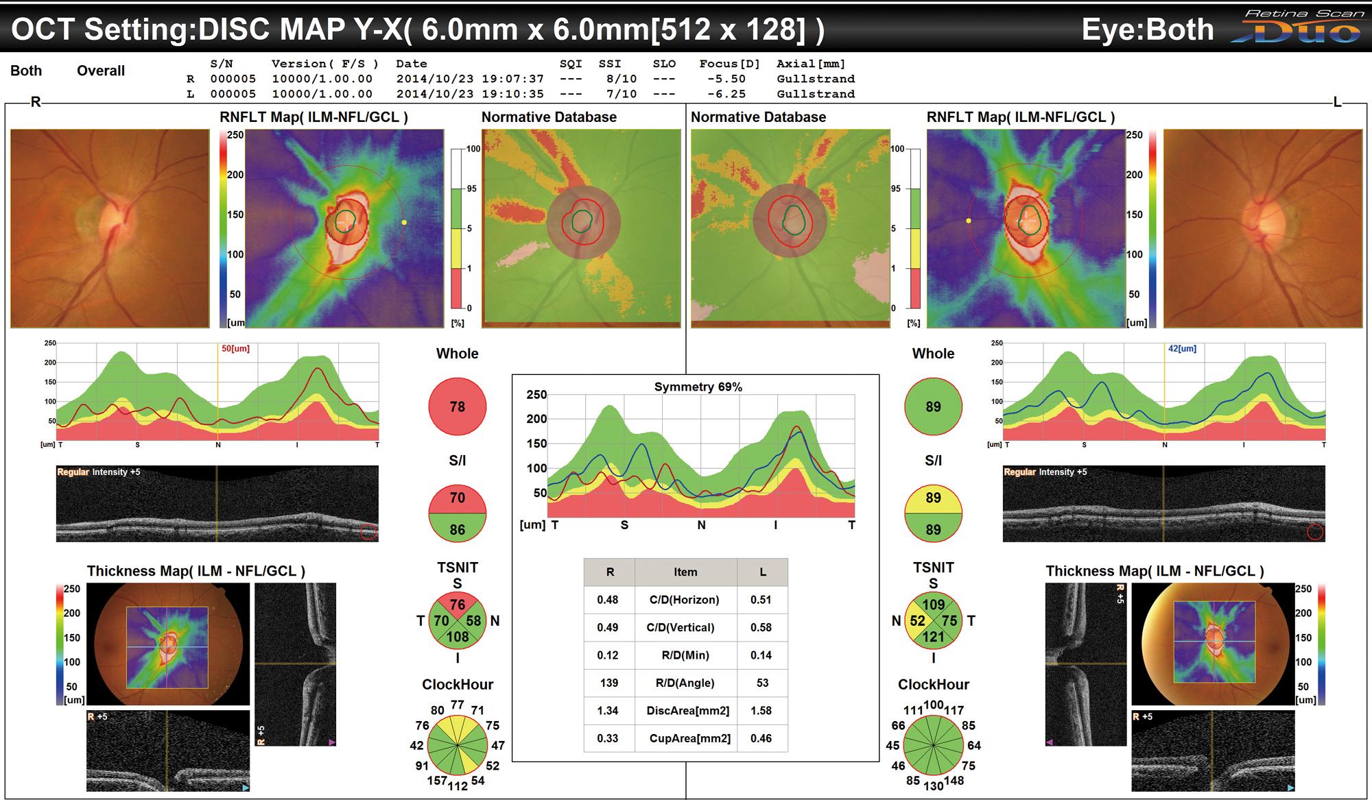
Once the data have been collected, they are analysed by the NAVIS-EX software platform, equipped with a viewer dedicated to OCT results and to the photos. Different map are displayed: thicknesses, global retina thickness and thickness of the complex of ganglionic cells, these thicknesses being compared with the normative 9x9 mm database, and the thickness of the nervous fibres of the optical nerve, in 6x6 mm. Also displayed are the OCT En-Face and all OCT sections, as well as the colour photos on which some measurements can be performed, such as the Disc and the Cup of papilla.

Legal mentions

These mentions are conforms to the French regulation and may vary depending on circumstances in each country.

Epaisseur du complexe des cellules ganglionnaires avec BDN sur 9x9mm (Glaucome)
Cartographie RNFL avec BDN sur 6x6mm (Glaucome)
Analyse OCT En-Face (drusen dans le cas d’une DMLA)
Représentation en 3D du Macula Map
Analyse du cube 3D ligne par ligne (Macula)
Line-by-line 3D cube analysis (macula)
Résultat du Macula Radial OD/OG
Résultat du Macula Line OD/OG

Standard accessories

  • External fixation point
  • Anterior segment adapter with additional front rest.
  • Cleaning air gun
  • Protective cover
  • Protective sheet for chinrest

Consumables

  • Protective sheet for chinrest
  • Télécharger les bonnes pratiques de nettoyage
  • Sales brochure
  • Auto fluorescence Clinical case book
THE OFFER OF NIDEK PRODUCTS

RS-330 additional products

Loading...

Mirante (SLO/OCT)

FIND MORE

NAVIS-EX Software

FIND MORE
PRODUCTS YOU MAY ALSO BE INTERESTED IN

In the same range

Loading...

RS-3000 Advance 2

FIND MORE

Mirante (SLO/OCT)

FIND MORE

Do you require more information?

You have a project? You want a quotation? You have questions about our products?
Feel free to ask your technical sales representative.

Contact us

The art of Eye Care

Reliability and
safety

NIDEK develops its top-of-the-range products to improve visual health through an approach based on strict criteria: safety, reliability, durability, continuous quality controls and certifications.

Technologies and
innovations

NIDEK meets technical challenges by keeping constantly informed of the innovations of eye imaging systems, using the expertise of professionals and the progresses of research.

Services and
guarantees

NIDEK commits itself to providing services to its customers, from the installation of an activity to the authorised training of teams, and to offering long-time measurable guarantees.
FIND MORE

Contact NIDEK

Contact us

Spécialiste des équipements et technologies au service de la santé visuelle, NIDEK SA accompagne et équipe les ophtalmologistes, orthoptistes, professionnels de l’optique et les établissements de santé.

Facebook Linkedin Youtube

KEY PRODUCTS

  • Mirante
  • TS 610
  • RT-Connect
  • Synetic

Services

  • Design office
  • Installation & Implementation
  • Trainings
  • ASS and repairing

Solutions

  • Optics
  • Ophthalmology
  • Orthoptics
  • Health care establishment

About us

  • Expertise and know-how
  • Our key dates
  • Certifications
  • Nidek in France and worldwide
  • Recruitment

© 2021 • NIDEK SA – All rights reserved

  • General Terms and Conditions
  • Protection des données personnelles
  • Legal notices
Gérer le consentement aux cookies
Nous utilisons des cookies pour optimiser notre site web et notre service.
Fonctionnel Always active
Le stockage ou l’accès technique est strictement nécessaire dans la finalité d’intérêt légitime de permettre l’utilisation d’un service spécifique explicitement demandé par l’abonné ou l’utilisateur, ou dans le seul but d’effectuer la transmission d’une communication sur un réseau de communications électroniques.
Préférences
Le stockage ou l’accès technique est nécessaire dans la finalité d’intérêt légitime de stocker des préférences qui ne sont pas demandées par l’abonné ou l’utilisateur.
Statistiques
Le stockage ou l’accès technique qui est utilisé exclusivement à des fins statistiques. Le stockage ou l’accès technique qui est utilisé exclusivement dans des finalités statistiques anonymes. En l’absence d’une assignation à comparaître, d’une conformité volontaire de la part de votre fournisseur d’accès à internet ou d’enregistrements supplémentaires provenant d’une tierce partie, les informations stockées ou extraites à cette seule fin ne peuvent généralement pas être utilisées pour vous identifier.
Marketing
Le stockage ou l’accès technique est nécessaire pour créer des profils d’utilisateurs afin d’envoyer des publicités, ou pour suivre l’utilisateur sur un site web ou sur plusieurs sites web ayant des finalités marketing similaires.
Manage options Manage services Manage {vendor_count} vendors Read more about these purposes
Préférences
{title} {title} {title}

Galerie

Suspicion de rétinite pigmentaire
Diabetic retinopathy
Behçet’s disease
Uvéite associée à la maladie de Vogt-Koyanagi-Harada
Glaucoma
Membrane épi-rétinienne associée à une suspicion de rétinite pigmentaire
Polypoid choroidal vasculopathy

Call me back

A problem was detected in the following Form. Submitting it could result in errors. Please contact the site administrator.

Télécharger votre brochure

Télécharger votre brochure

Télécharger votre brochure

Télécharger votre brochure

Télécharger votre brochure

Télécharger votre brochure

Télécharger votre brochure

Télécharger votre brochure

Télécharger votre brochure

Télécharger votre brochure

Télécharger votre brochure

Télécharger votre brochure

Télécharger votre brochure

Télécharger votre brochure

Télécharger votre brochure

Télécharger votre brochure

Télécharger votre brochure

Télécharger votre brochure

Télécharger votre brochure

Télécharger votre brochure

Télécharger votre brochure

Télécharger votre brochure

Télécharger votre brochure

Télécharger votre brochure

Ce site est réservé aux professionnels de santé

Sortir
Je suis professionnel de santé