• 01 49 80 97 97
  • Contact us
  • Boutique NIDEK

Efficient After Sales Service

At your disposal whatever the situation

Made in France

Our consultation units

Our experts at your disposal

60 experts over the territory

  • Lasers
        • Lasers YAG and SLT
          • YC-200 S Plus (YAG/SLT)
          • YC-200 (YAG)
        • Photocoagulator lasers
          • YLC-500 / YLC 500 VIXI
          • GYC-500 / GYC 500 VIXI
  • Retina diagnosis
        • Slit lamps
          • SL-9900/SL-9900 Zoom
          • SL-9800
          • SL-250
        • Multimodal platforms
          • Mirante (SLO/OCT)
          • Mirante SLO (SLO)
        • Fundus cameras
          • AFC-330
          • Retina Scan Duo 2
          • Mirante SLO (SLO)
          • Mirante (SLO/OCT)
        • Microperimeters
          • MP-3
          • MP-3S
        • Angiographs
          • Mirante (SLO/OCT)
          • Mirante SLO (SLO)
        • Software
          • NAVIS-EX Software
        • OCT
          • RS-1
          • Retina Scan Duo 2
          • Mirante (SLO/OCT)
        • Ultrasound scanners
          • US-4000/4000-P (Echo A+B)
          • US-500 / 500-P (Echo A)
  • Anterior segment
        • Slit lamps
          • SL-9900/SL-9900 Zoom
          • SL-9800
          • SL-250
        • Aberrometers - Topographs
          • OPD-Scan III
        • Optical biometers
          • AL-Scan
          • AL-Scan M
        • Tonometers
          • TONOREF III
          • NT-1e / NT-1 / NT-1p
        • Specular microscope
          • CEM-530
        • Goniolenses
          • GS-1
  • Refraction
        • Pupillometers
          • PM-700
        • Afficheurs de tests
          • CP-9
          • SC-1600 Pola/SC-1600
        • Itinerant optician case
          • Itinerant optician case
        • Lensometers
          • LM 1800 P – PD
          • LM7 – 7P
        • Auto refractometers
          • AR-1/ARK-1
          • AR-1a/ARK-1a
          • AR-1s/ARK-1s
          • AR-F/ARK-F
          • HandyRef/HandyRef-K
          • TONOREF III
        • Refractors
          • RT-3100
          • TS-310
          • RT-6100
          • TS-610
          • Logiciel RT-Connect
        • Système de réfraction assisté
          • FARS (Fully Assisted Refraction System)
  • Consultation units
        • "Excellence" units
          • Synetic
          • OT-6400
        • "Performance" units
          • Affinity
          • OT-4200
        • "Compact" units
          • ONE-i
          • Lypop
          • COS-6100
        • Lifting tables and examination swivel chair
          • Consultation armchairs and stools
          • Tables à élévation Up
  • Optical workshop
        • Automatic edges
          • ME-1500
          • LEXCE Trend 8D
          • LEXCE Trend D
          • LEXCE Trend DBT
          • SE9090 Supra
        • Lensometers
          • LM 1800 P – PD
          • LM7 – 7P
        • Centering-blocking devices
          • ICE-1500
          • ICE- 1
        • Shape plotters
          • LT-980
          • LETR-3 Traceur LEXCE Trend
        • Green products
          • LFU-220
          • LED-200 Plus
        • Colonne de centrage : Invision Fit AI
  • NIDEK
        • Our services
          • Design office
          • Installation & Implementation
          • Trainings
          • ASS and repairing
          • Environement
          • Showroom NIDEK
        • Our solutions
          • Optics
          • Ophthalmology
          • Orthoptics
          • Health care establishment
        • About us
          • Expertise and know-how
          • Certifications
          • NIDEK in France
          • NIDEK worldwide
          • Recruitment
        • News
          • Blog
          • Évènements
  • Our shop
  • fr_FR
  • en_GB

Homepage > Angiography

Angiographs

IMAGERIE vasculaire DE LA RÉTINE
Our products
Benefits
Getting equipped
DYNAMIC OBSERVATION

Diagnosis and therapeutic orientation

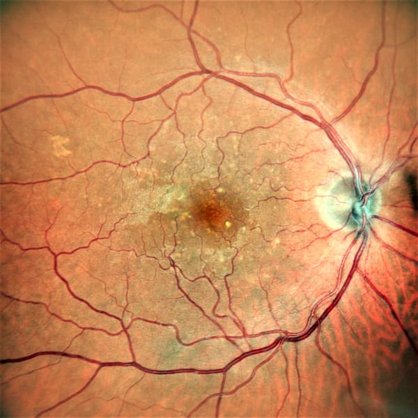
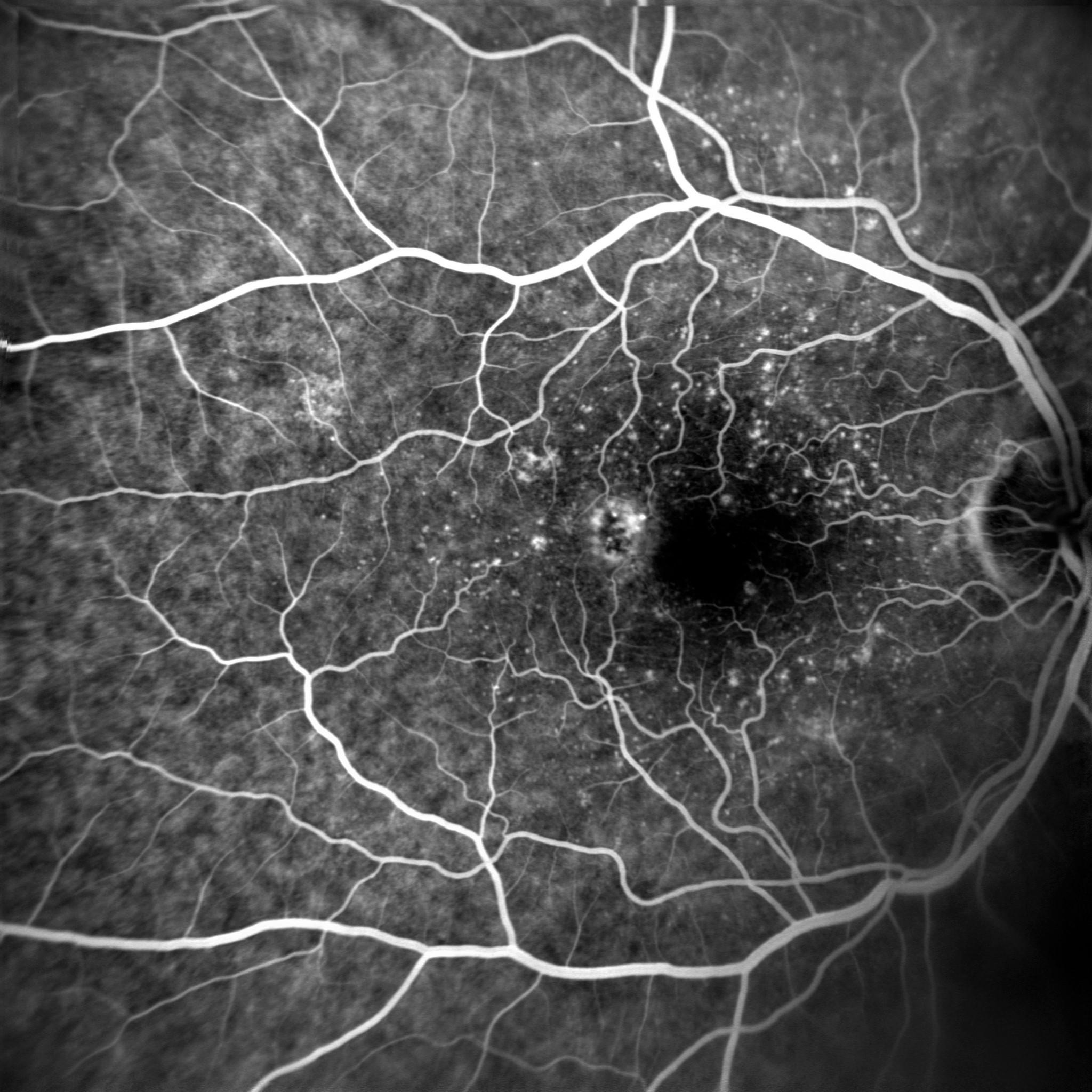
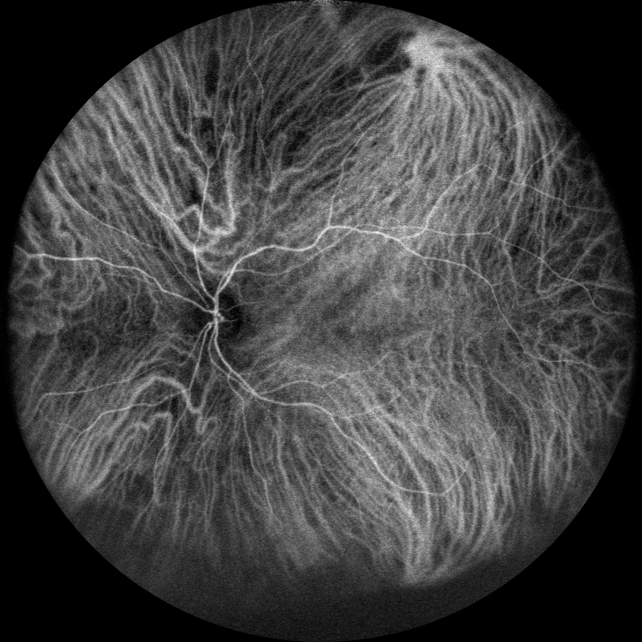
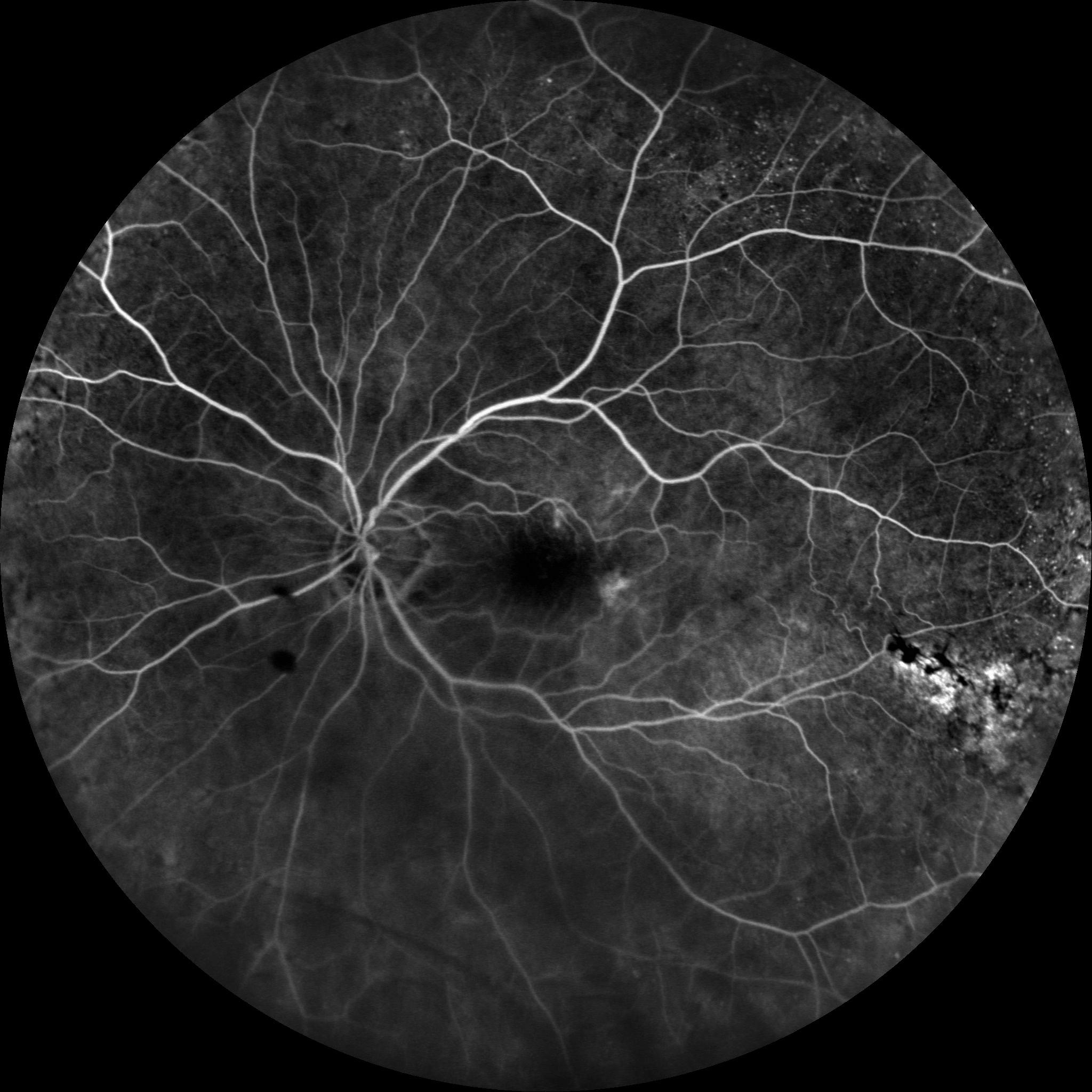
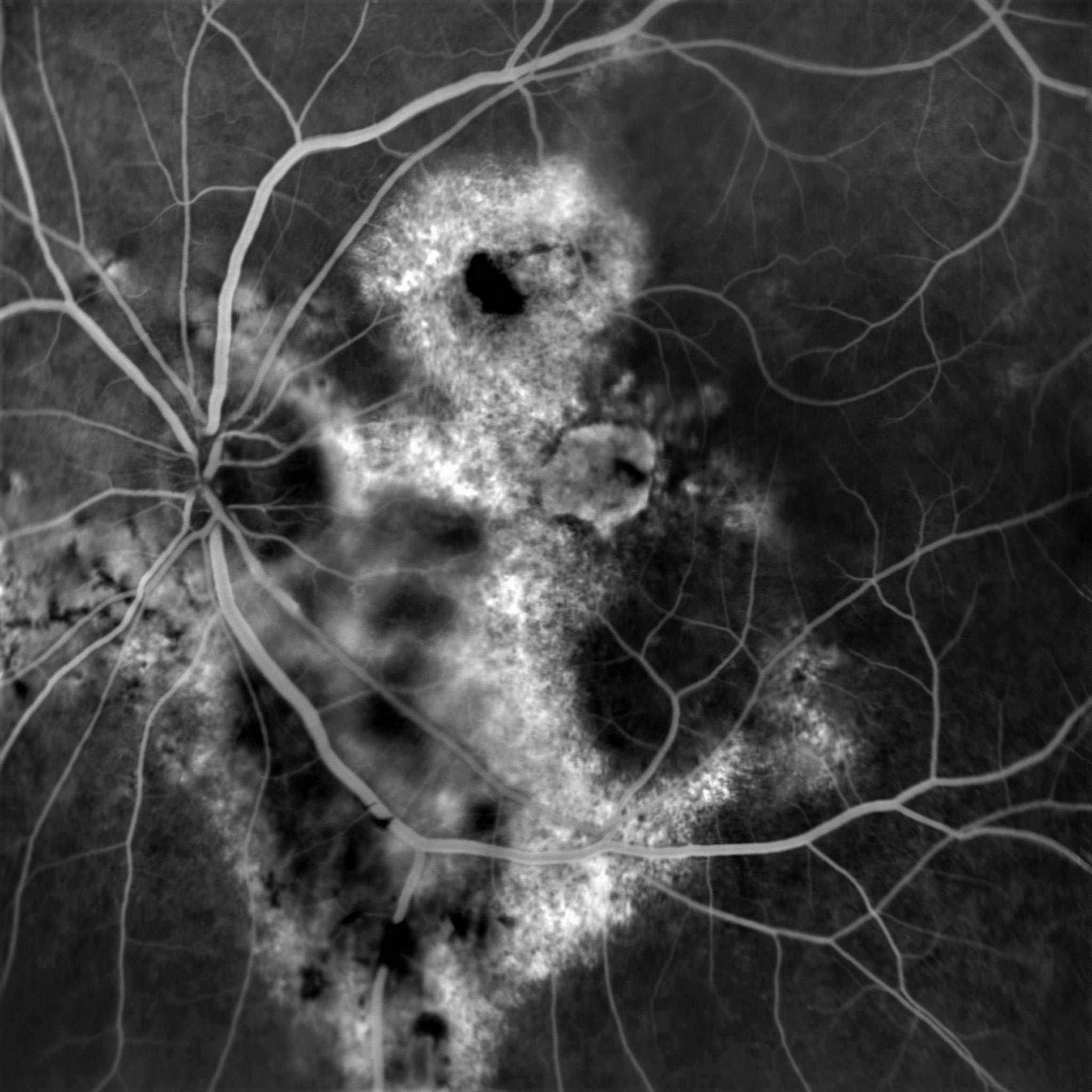
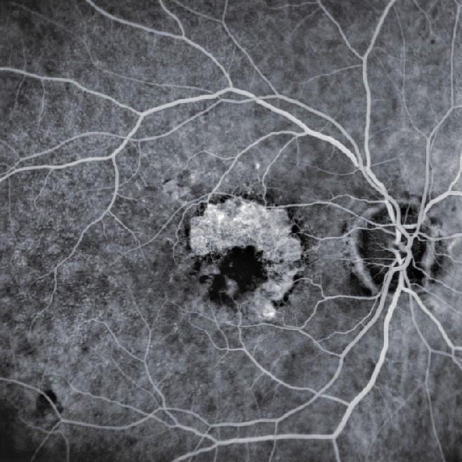
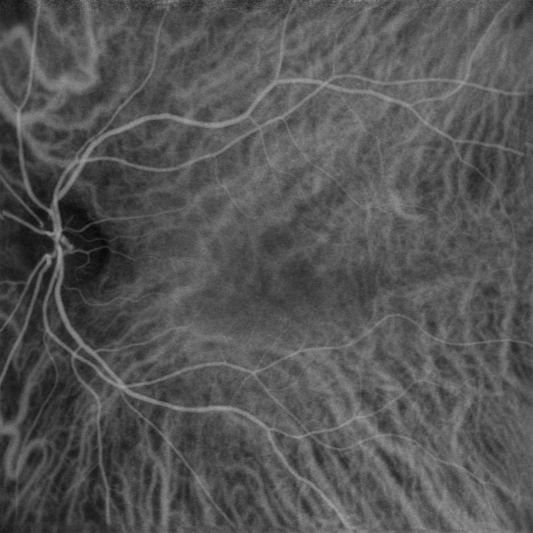
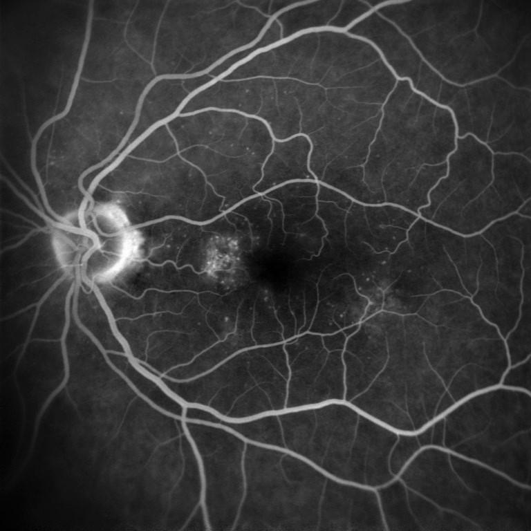
Retinal angiography is a dynamic diagnostic examination allowing the visualisation of the vascular system of the retina and detection of abnormalities that are not always visible with other fundus imaging methods.

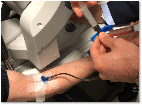
Performed using a fluorescent contrast product (colour) injected into the patient’s blood circulation, it is therefore seen as invasive and requires a convenient equipment to perform the injection and the resuscitation in case of allergic reaction.

L’angiographie trouve son utilité pour le diagnostic et le suivi de pathologies vasculaires telles que la rétinopathie diabétique, la AMD, les uvéites postérieures…

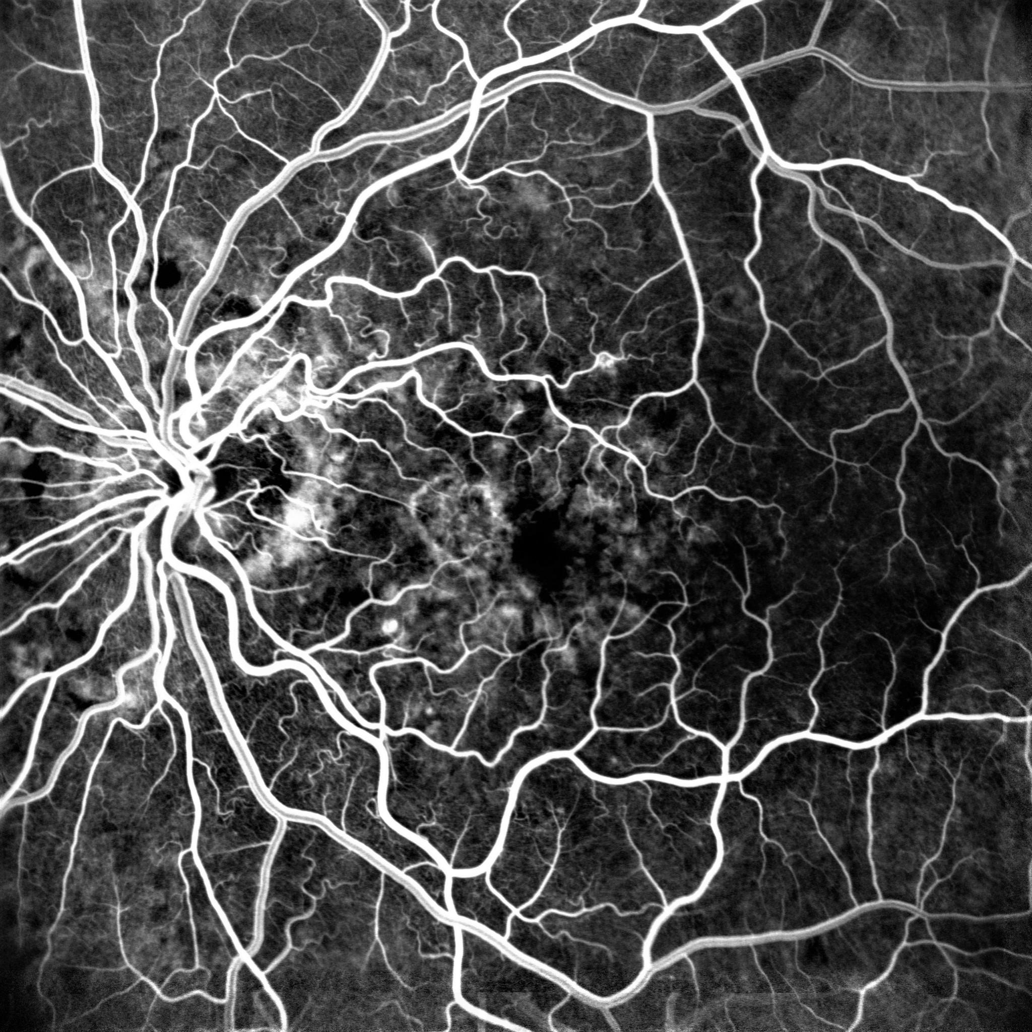
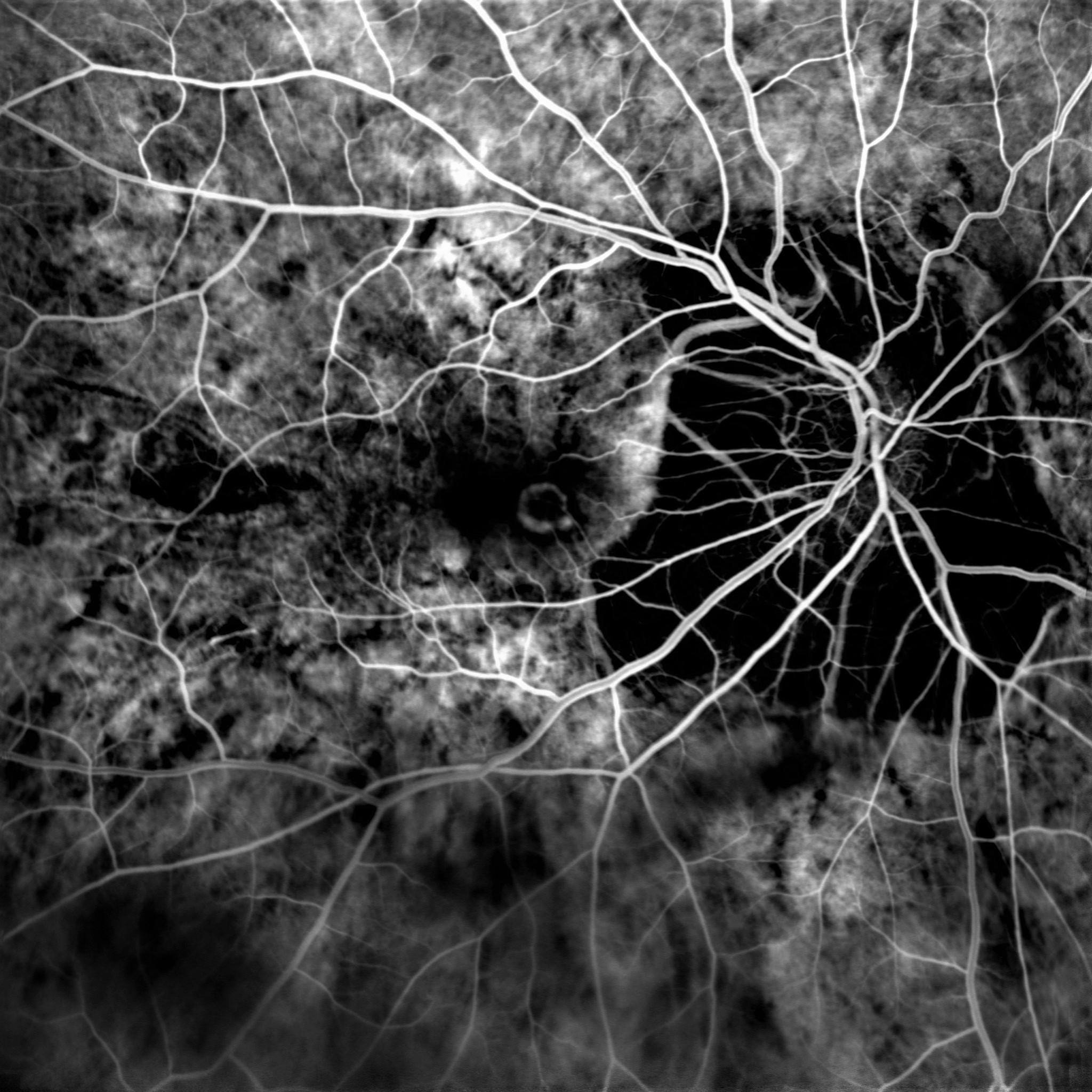
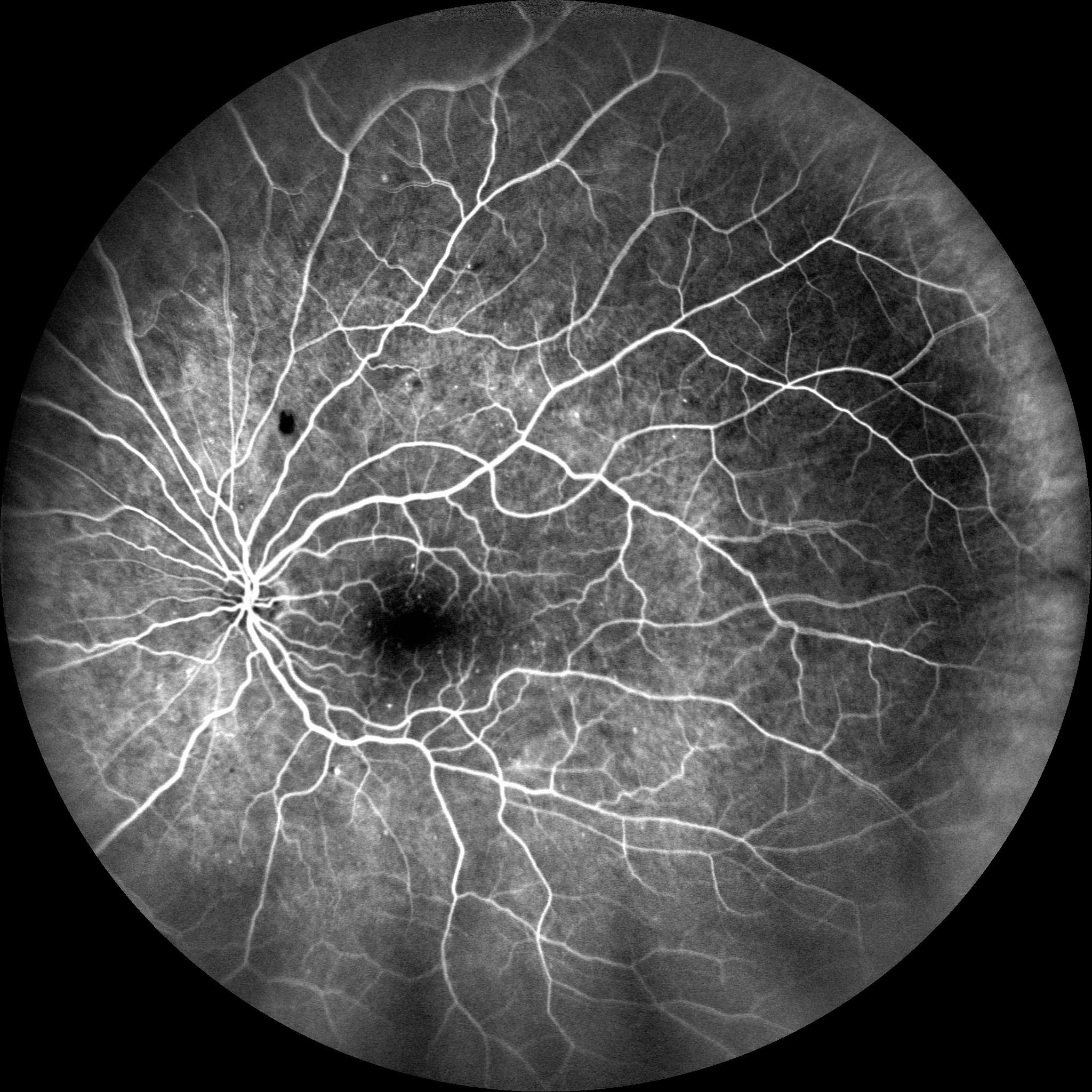
Depending on the depth of the tissue to examine and on the pathology, two different contrast products can be used. So they define two types of angiography, the fluorescein one (FA) and the indocyanine green one (ICG).

Current angiographs, often equippedwith a confocal SLO laser system.The current angiographs, often equipped with an SLO confocal system, able to precisely focus on retina, make possible performing the fluorescein angiography only, the indocyanine green angiography only or sometimes both angiographies at the same time.

NIDEK PRODUCTS

Our angiography solutions

Loading...

Mirante SLO (SLO)

Mirante is a large-field multimodal-imaging platform designed to perform fundus imaging. It combines OCT and SLO technologies thus grouping routine imaging to detect and identify retinal-choroidal pathologies. 

FIND MORE

Mirante (SLO/OCT)

Mirante is a large-field multimodal-imaging platform designed to perform fundus imaging. It combines OCT and SLO technologies thus grouping routine imaging to detect and identify retinal-choroidal pathologies. Using an additional lens, performing an analysis of sections of the anterior segment is also possible.

FIND MORE
NIDEK EXPERTISE

Techniques used

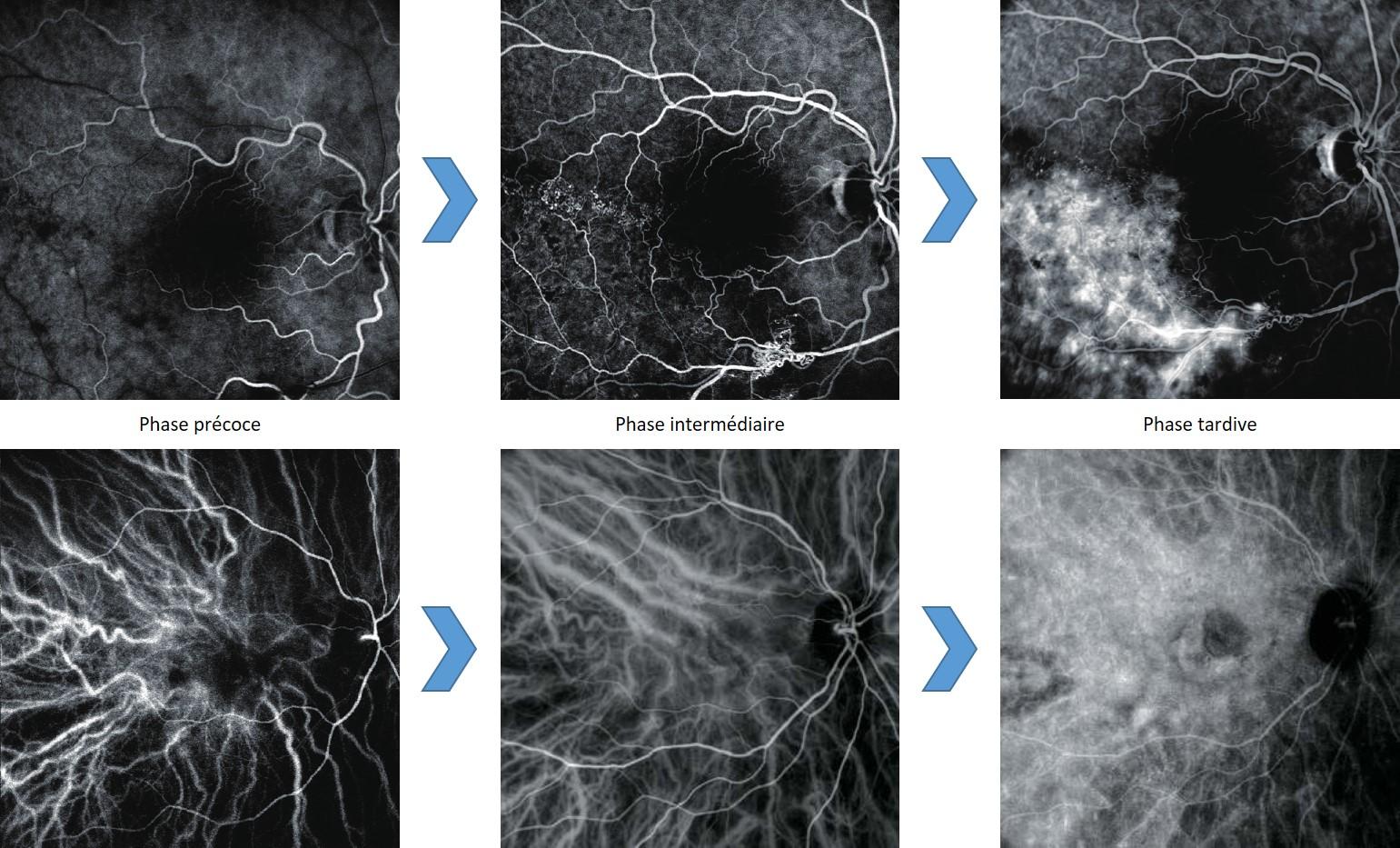
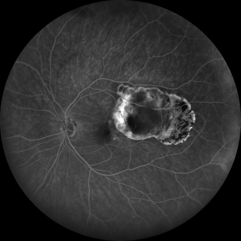
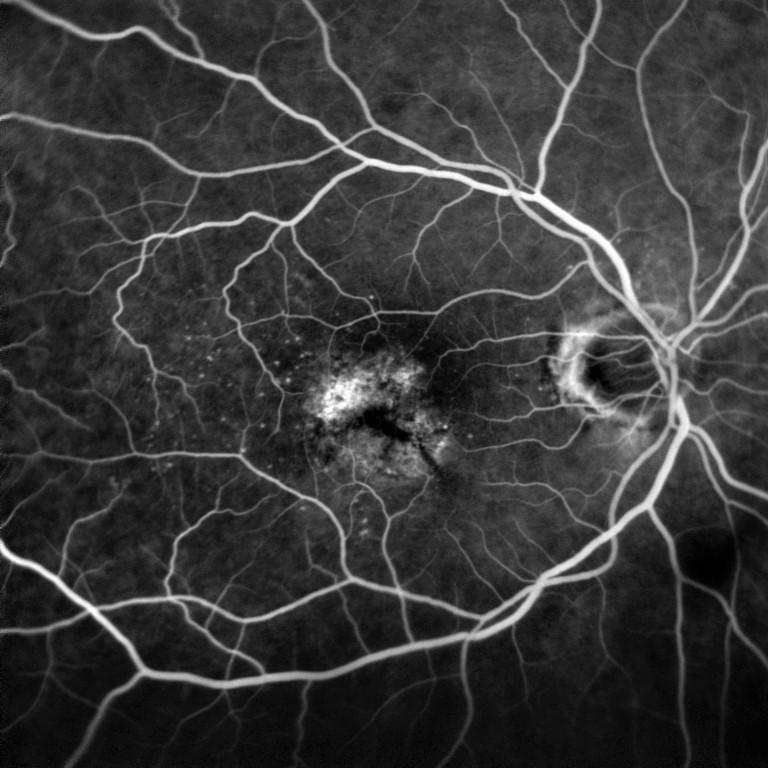
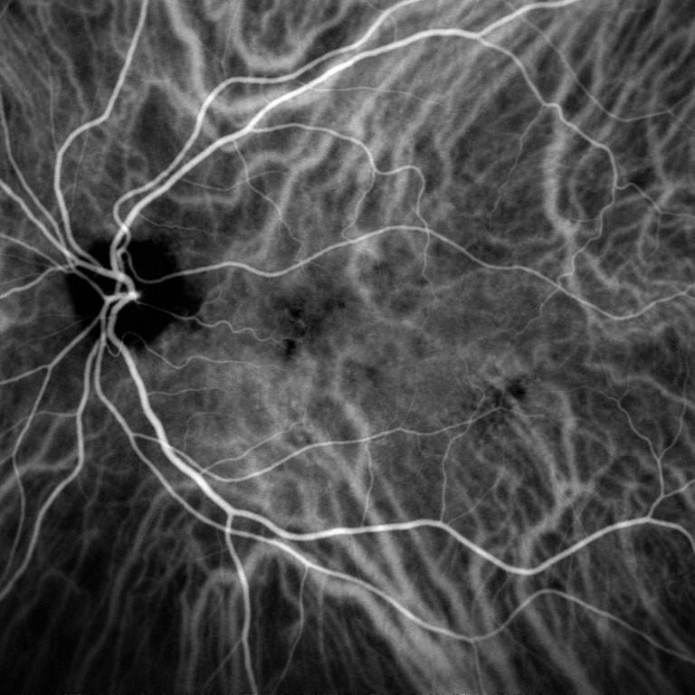
The injection angiography is the only examination making possible viewing fundus vascularisation in a dynamic way. It is then possible to analyse early, intermediate and late phases of retinal and choroidal blood circulations.

Angiography is often performed using an SLO angiograph, as this equipment can make a precise focus on retina. The purpose is detecting vascularisation anomalies, such as:

  • Aneurysms
  • Leaks
  • Oedemas
  • Ischemia 
  • AMD
  • Diabetic retinopathy
  • Vascular occlusions and tumours, etc.

The examination is performed using a contrast product injected into the patient’s blood circulation.

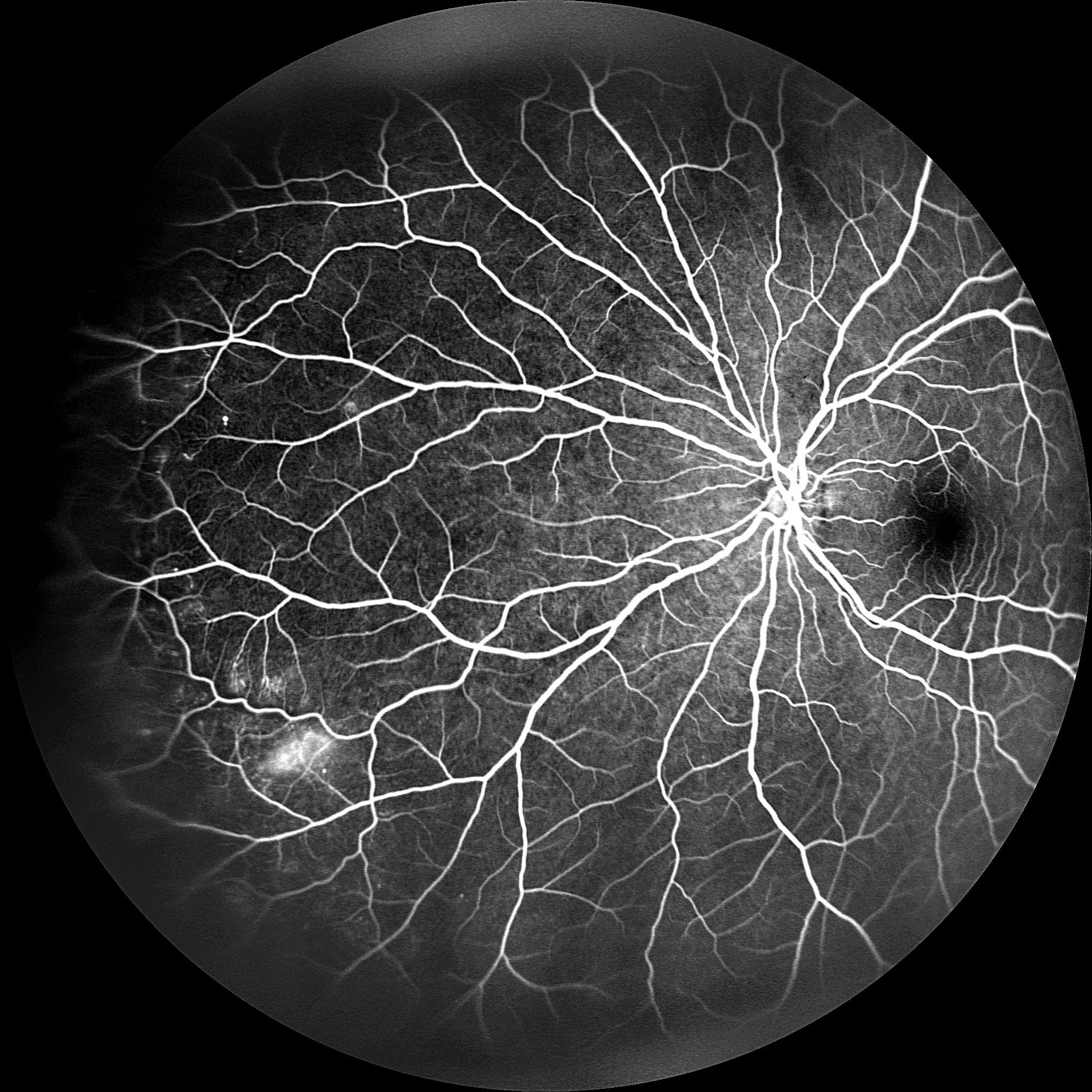
During the examination, the angiograph illuminates the fundus with a specific wavelength to stimulate the colour to make it visible. Angiography images then only show the colour circulation into the vascular system, thus matching the blood behaviour into the vessels.

We use angiography to make a precise diagnosis, to propose an adapted treatment and to follow-up the evolution of the patient’s pathology through a control. When a complete analysis of the structure/function of the retina is required, the angiography is always used in addition to the OCT.

This imaging technique is different from the OCT-Angiography, which is non invasive, as it derives from the OCT (scanner of the retina). This last technique remains static and for the moment its acquisition field is limited.

Pathologies that can be detected using an angiograph

We can use the result to control the condition of the retinal and choroidal vascular system, affected in many eye pathologies:

Image courtesy: Dr Djaborouti (Puilboreau, France), hospital Luigi Sacco (University of Milan, Italy), hospital Lariboisière (Paris, France).

FA OR ICG ANGIOGRAPHY

The different types of angiography

According to the pathology and the vascularisation to observe, you can use different contrast products. We know the fluorescein one and the indocyanine green one.

Fluorescein angiography (FA)

Fluorescein angiography is the most commonly used. It is particularly used before a session of laser photocoagulation. It is used to perform the analysis of the retinal vascularisation of surface layers. The examination takes only ten minutes.

Indocyanine green angiography (ICG)

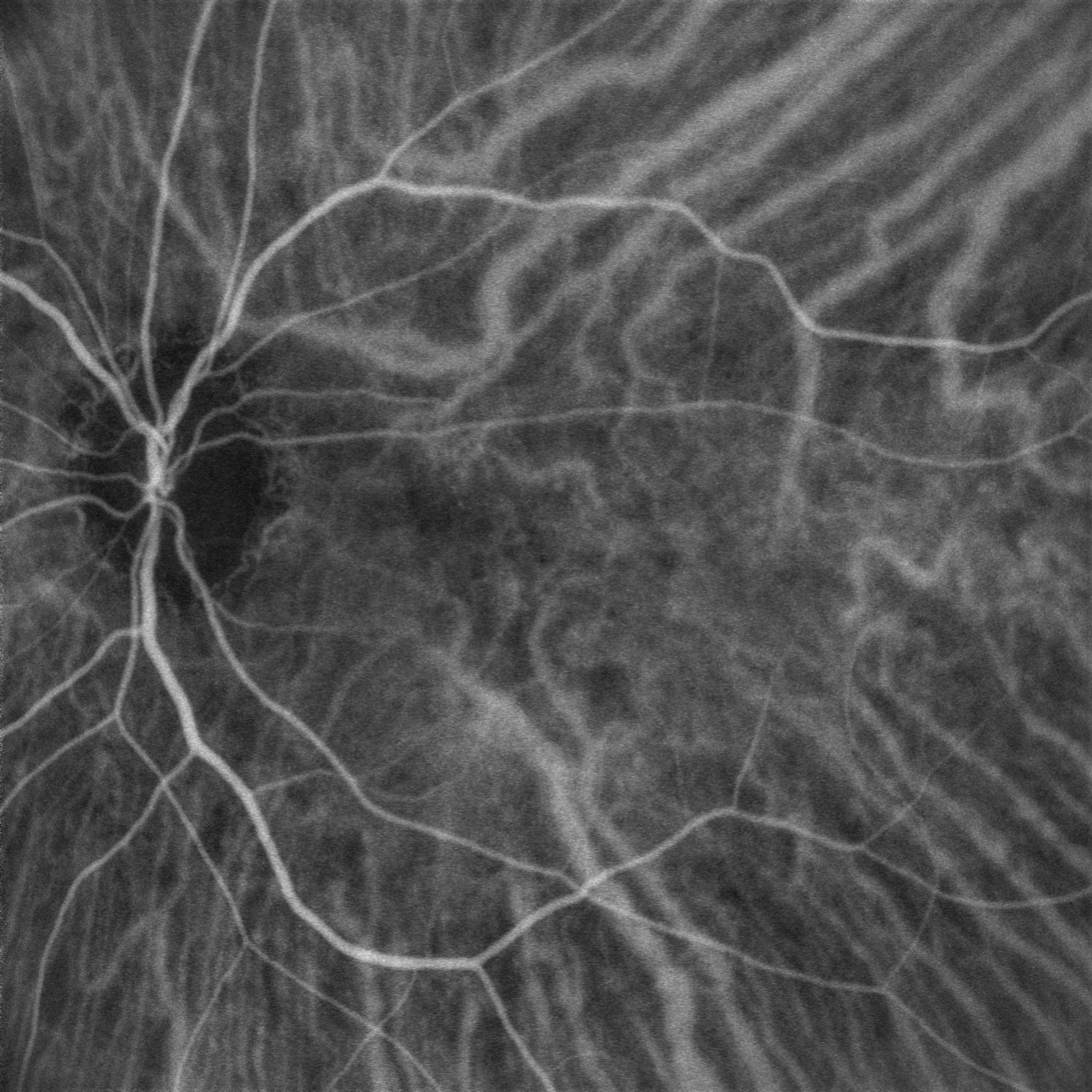
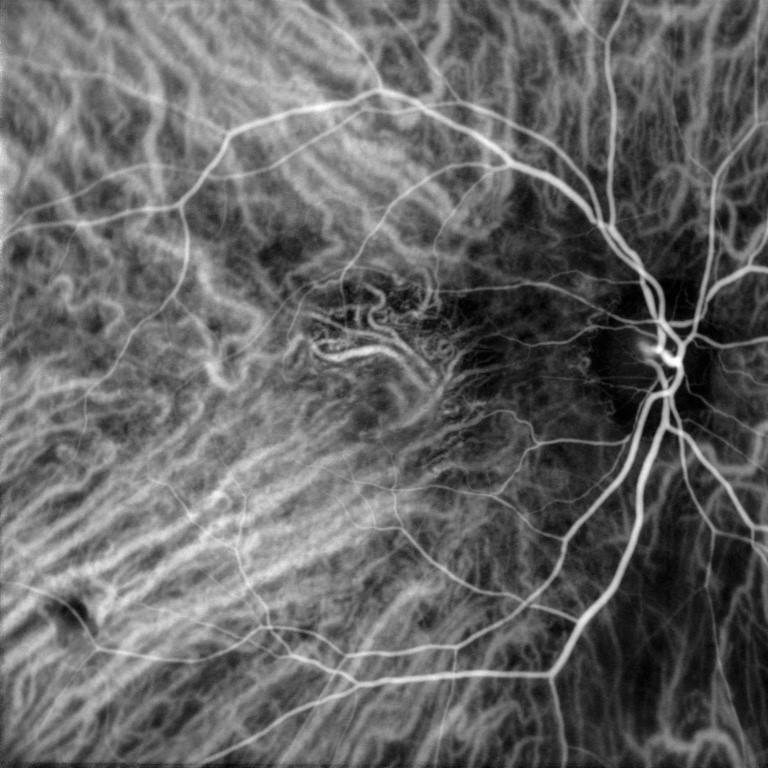
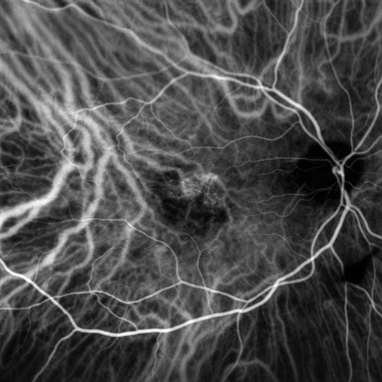
The indocyanine green angiography is less used but it is the only method making possible viewing the vascularisation of deep layers including the choroidal layer. The examination is performed under infrared light and can last up to 30 minutes.

Benefits of angiography

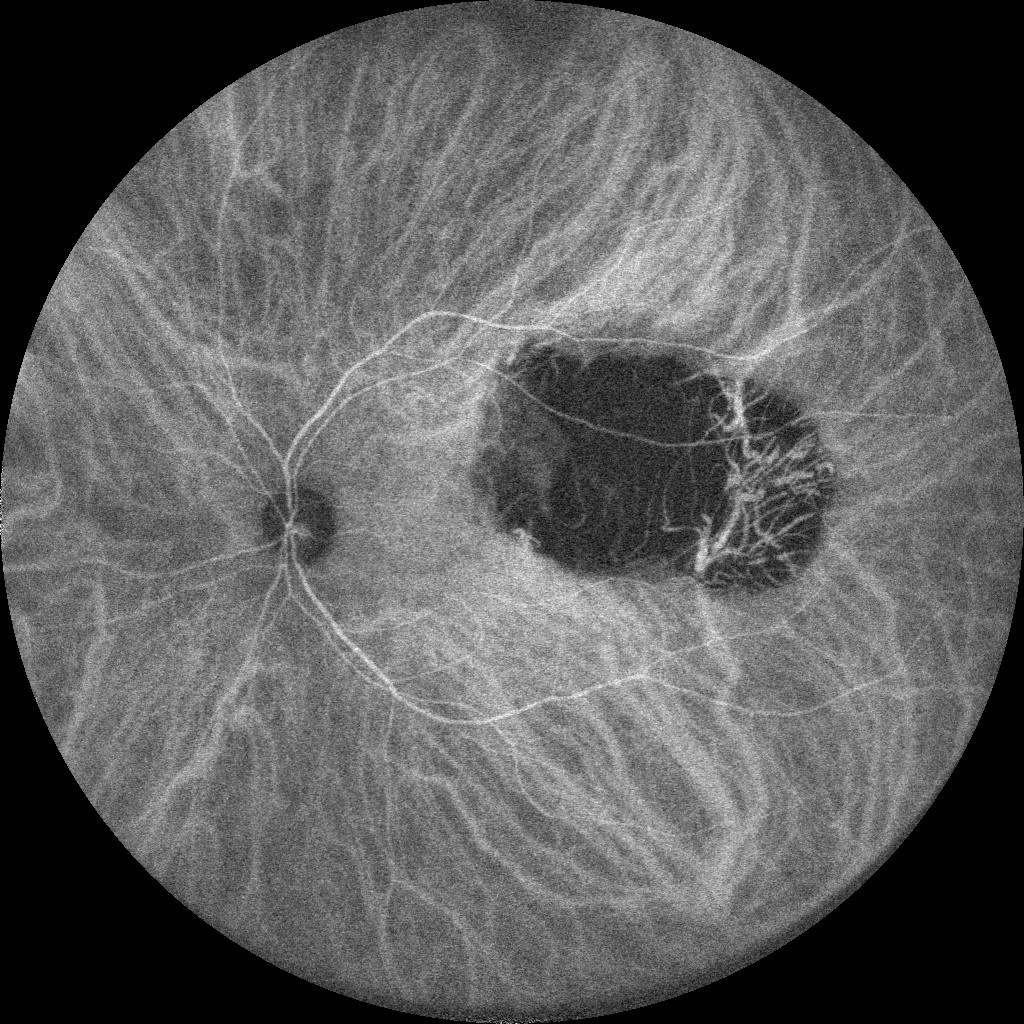
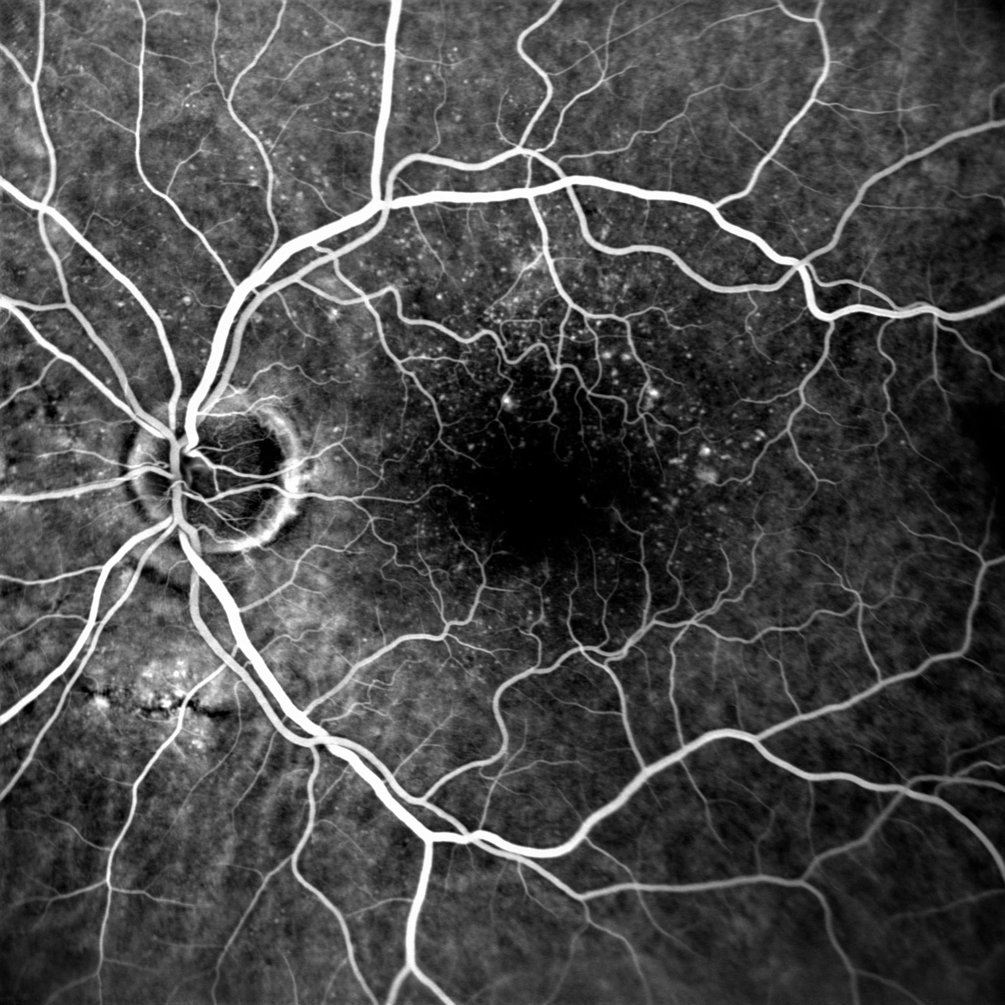
The angiography is the only technique realising a dynamic observation of fundus vascularisation. Depending on the lens used, the analysis zone can be extended to make possible detecting some early vascular anomalies.

  • Dynamic analysis

Dynamic analysis of retinal and choroidal vascularisation

  • Extended acquisition

Visualisation du fond d’œil jusqu’à l’extrême périphérie grâce au grand champ

  • Assistance to diagnosis

The vascular analysis completes the structural analysis performed using fundus photography and the OCT to confirm and refine the diagnosis.

Do you want to purchase an angiograph?

You have a project? You want a quotation? You have questions about our products? Feel free to ask your technical sales representative.

Contact us

The Art of Eye Care

Reliability and
safety

NIDEK develops its top-of-the-range products to improve visual health through an approach based on strict criteria: safety, reliability, durability, continuous quality controls and certifications.

Technologies and
innovations

NIDEK meets technical challenges by keeping constantly informed of the innovations of eye imaging systems, using the expertise of professionals and the progresses of research.

Services and
guarantees

NIDEK commits itself to providing services to its customers, from the installation of an activity to the authorised training of teams, and to offering long-time measurable guarantees.

FIND MORE
Le magazine

L'actualité NIDEK

Notre actualité

Les OCT Nidek référencés par l’URPS

Les OCT Nidek référencés par l’URPS : Une opportunité pour les professionnels de santé visuelle

Lire la suite »

🎉 NIDEK à la SFO 2025 : notre gamme médicale à l’honneur !

🎉 NIDEK à la SFO 2025 : notre gamme médicale à l’honneur ! Du 10

Lire la suite »

NIDEK accompagne la formation des assistants médicaux en ophtalmologie avec Keyce Santé

Chez NIDEK, nous sommes convaincus que l’innovation ne se limite pas aux équipements : elle

Lire la suite »

Retour sur la visite de M. Ozawa, président de NIDEK, en France

Le 11 février, NIDEK SA France a eu l’honneur d’accueillir M. Ozawa, président de NIDEK,

Lire la suite »
Découvrir le blog
Nos événements

🎉 NIDEK à la SFO 2025 : notre gamme médicale à l’honneur !

🎉 NIDEK à la SFO 2025 : notre gamme médicale à l’honneur ! Du 10

Lire la suite »

Retour sur la visite de M. Ozawa, président de NIDEK, en France

Le 11 février, NIDEK SA France a eu l’honneur d’accueillir M. Ozawa, président de NIDEK,

Lire la suite »

Nidek et MEDADOM renouvellent leur partenariat pour leur dispositif innovant de téléophtalmologie

Communiqué de presse Nidek et MEDADOM renouvellent leur partenariat pour leur dispositif innovant de téléophtalmologie

Lire la suite »

NIDEK SA à la Journée Nationale de Téléophtalmologie 2024

Nous sommes ravis d’annoncer la participation de NIDEK SA à la Journée Nationale de Téléophtalmologie

Lire la suite »
Tous nos événements

Contact NIDEK

Contact us

Spécialiste des équipements et technologies au service de la santé visuelle, NIDEK SA accompagne et équipe les ophtalmologistes, orthoptistes, professionnels de l’optique et les établissements de santé.

Facebook Linkedin Youtube

KEY PRODUCTS

  • Mirante
  • TS 610
  • RT-Connect
  • Synetic

Services

  • Design office
  • Installation & Implementation
  • Trainings
  • ASS and repairing

Solutions

  • Optics
  • Ophthalmology
  • Orthoptics
  • Health care establishment

About us

  • Expertise and know-how
  • Our key dates
  • Certifications
  • Nidek in France and worldwide
  • Recruitment

© 2021 • NIDEK SA – All rights reserved

  • General Terms and Conditions
  • Protection des données personnelles
  • Legal notices
Gérer le consentement aux cookies
Nous utilisons des cookies pour optimiser notre site web et notre service.
Fonctionnel Always active
Le stockage ou l’accès technique est strictement nécessaire dans la finalité d’intérêt légitime de permettre l’utilisation d’un service spécifique explicitement demandé par l’abonné ou l’utilisateur, ou dans le seul but d’effectuer la transmission d’une communication sur un réseau de communications électroniques.
Préférences
Le stockage ou l’accès technique est nécessaire dans la finalité d’intérêt légitime de stocker des préférences qui ne sont pas demandées par l’abonné ou l’utilisateur.
Statistiques
Le stockage ou l’accès technique qui est utilisé exclusivement à des fins statistiques. Le stockage ou l’accès technique qui est utilisé exclusivement dans des finalités statistiques anonymes. En l’absence d’une assignation à comparaître, d’une conformité volontaire de la part de votre fournisseur d’accès à internet ou d’enregistrements supplémentaires provenant d’une tierce partie, les informations stockées ou extraites à cette seule fin ne peuvent généralement pas être utilisées pour vous identifier.
Marketing
Le stockage ou l’accès technique est nécessaire pour créer des profils d’utilisateurs afin d’envoyer des publicités, ou pour suivre l’utilisateur sur un site web ou sur plusieurs sites web ayant des finalités marketing similaires.
Manage options Manage services Manage {vendor_count} vendors Read more about these purposes
Préférences
{title} {title} {title}

Télécharger votre brochure

Télécharger votre brochure

Télécharger votre brochure

Télécharger votre brochure

Télécharger votre brochure

Télécharger votre brochure

Télécharger votre brochure

Télécharger votre brochure

Télécharger votre brochure

Télécharger votre brochure

Télécharger votre brochure

Télécharger votre brochure

Télécharger votre brochure

Télécharger votre brochure

Télécharger votre brochure

Télécharger votre brochure

Télécharger votre brochure

Télécharger votre brochure

Télécharger votre brochure

Télécharger votre brochure

Télécharger votre brochure

Télécharger votre brochure

Télécharger votre brochure

Télécharger votre brochure

Ce site est réservé aux professionnels de santé

Sortir
Je suis professionnel de santé