Efficient After Sales Service
At your disposal whatever the situation
Made in France
Our consultation units
Our experts at your disposal
60 experts over the territory
Efficient After Sales Service
At your disposal whatever the situation
Made in France
Our consultation units
Our experts at your disposal
60 experts over the territory
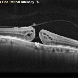
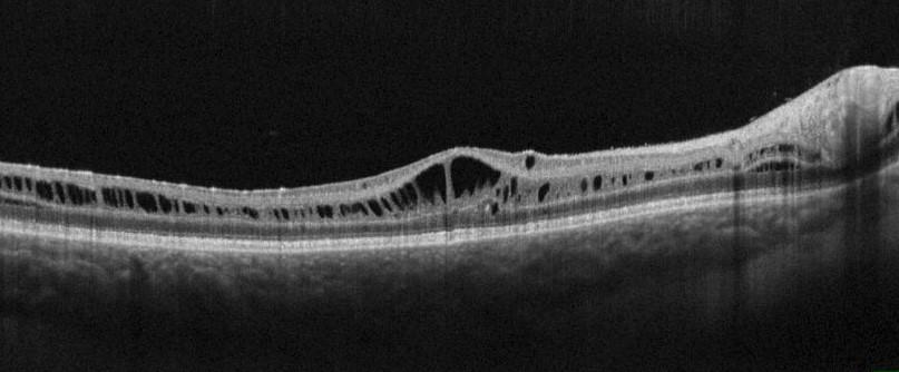
The OCT (Optical Coherence Tomography) has become a compulsory examination of retina. Thanks to its non-invasive acquisition method, in real time and with a micron-size resolution of the micrometre, it is used for the detection, the in-deep search of diagnosis and the patient’s following-up. This instrument scans the fundus using a laser beam and rebuilds sections and volumes to enable the detailed analysis of the entire retina and choroid layers. Therefore it is a precious tool for the detection and following-up of retinal-choroidal and papilla pathologies, such as AMD, retinopathies and maculopathies, glaucomas or genetic pathologies.
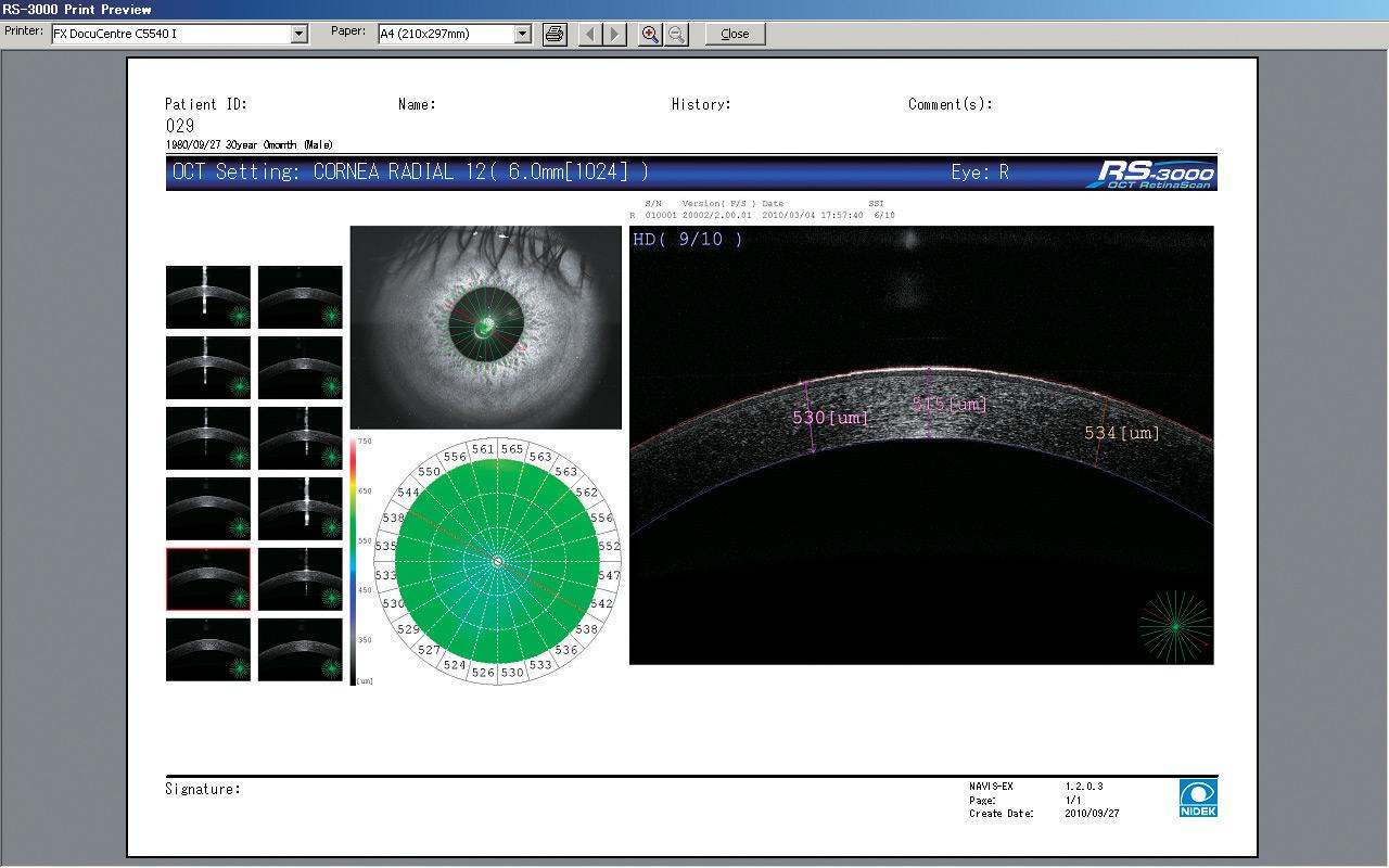
Thanks to an adaptor that takes the focal plane of the device to the front of the eye, or thanks to a specific OCT of the anterior segment, it is also possible to get sections of the anterior part of the eye, from the cornea to the crystalline lens. This use provides information about cornea structure and the opening of the iridocorneal angle, as this value is particularly controlled for patients suffering with glaucoma. OCT’s are always backed by a fundus imaging system and used to control the retina vasculature using an OCT-angiography.
NIDEK develops all the technologies related to both OCT and OCT-A.
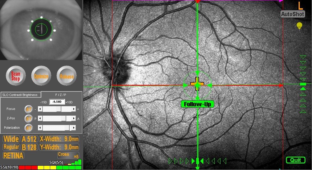
The Retina Scan Duo 2, combined fundus and autofluorescence OCT, is equipped with a 3D eye tracking system and automation to simplify measurement.
The RS-3000 Advance 2 is an OCT Spectral Domain Expert equipped with technology SLO and a function OCT-Angiography. These advanced functions make it a powerful diagnosis tool of retinal-choroidal pathologies. Thanks to an additional lens, it also makes possible analysing sections of the anterior segment.
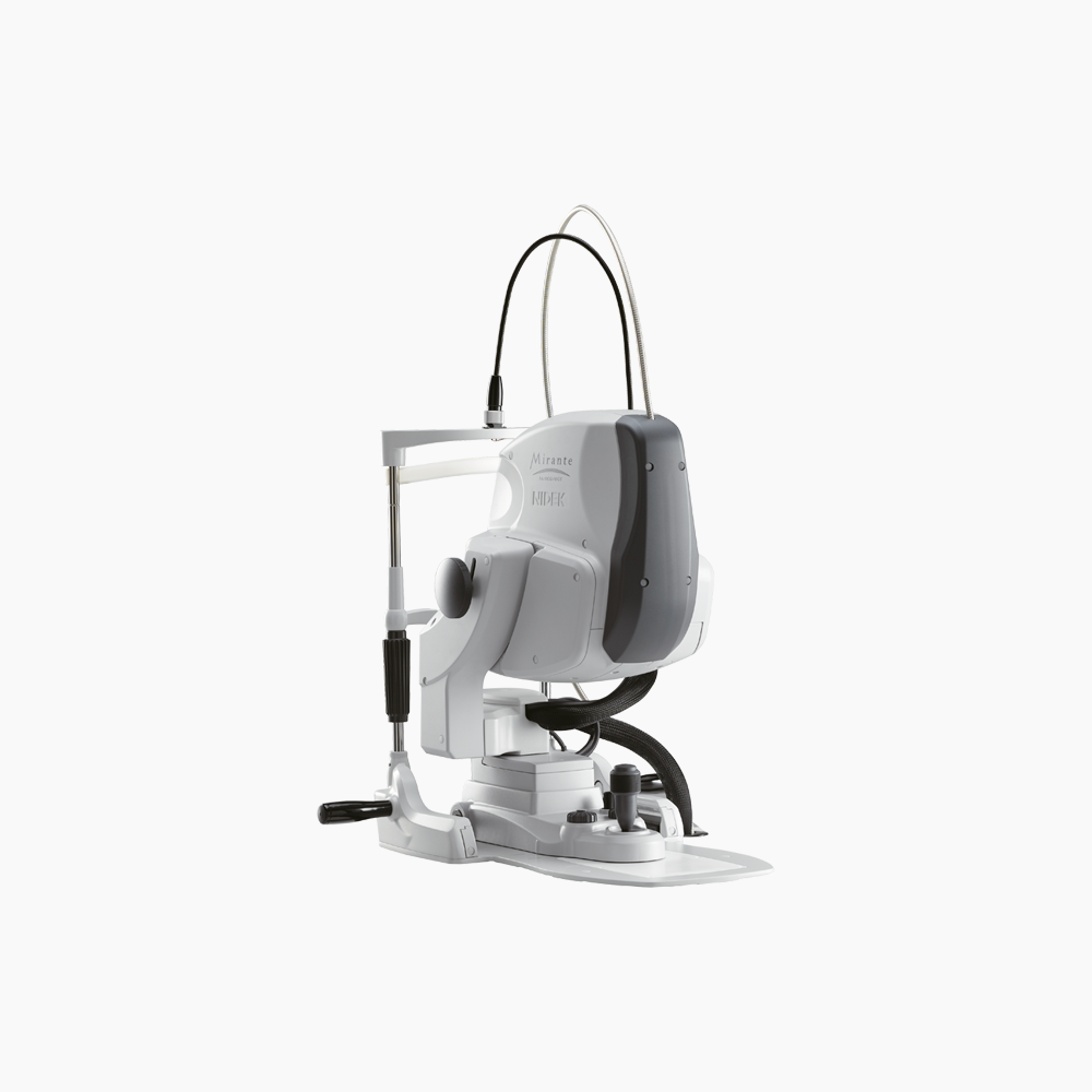
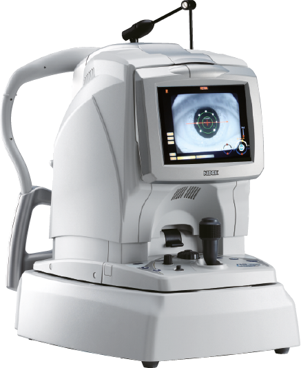
Mirante is a large-field multimodal-imaging platform designed to perform fundus imaging. It combines OCT and SLO technologies thus grouping routine imaging to detect and identify retinal-choroidal pathologies. Using an additional lens, performing an analysis of sections of the anterior segment is also possible.
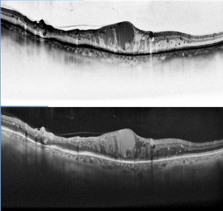
The OCT works using the bidirectional motion of an infrared laser source to scan the fundus. As for an echography, OCT sensors detect the echo of the signal reflected by the different arrangements of retina cellular layers.
This technique, originally based upon the interferometry principle, displays all cellular layers with micron-size resolution. The OCT scanning is positioned and relocated on the retina using another imaging system of the fundus (infrared or SLO), that impacts the tracking quality of the retina, to correctly position the acquisitions and the tracking examinations (sections collected in the same conditions).
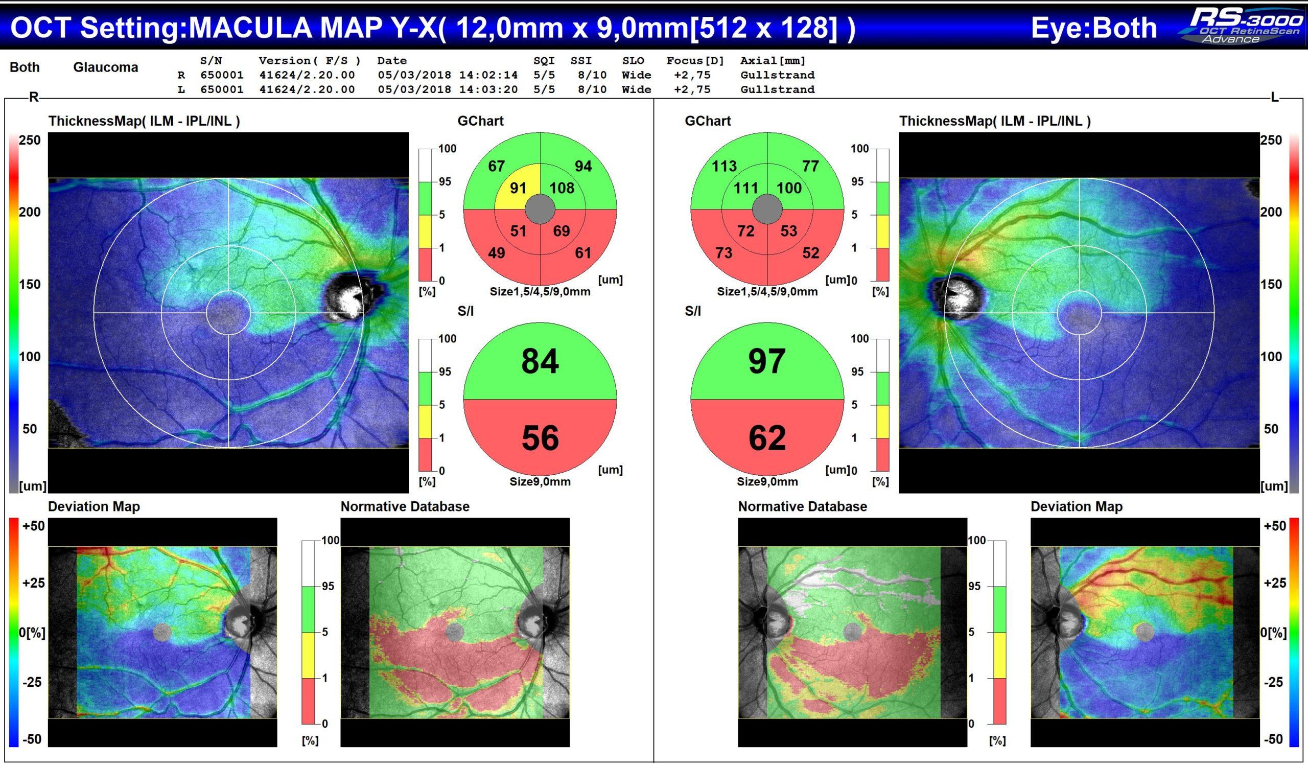
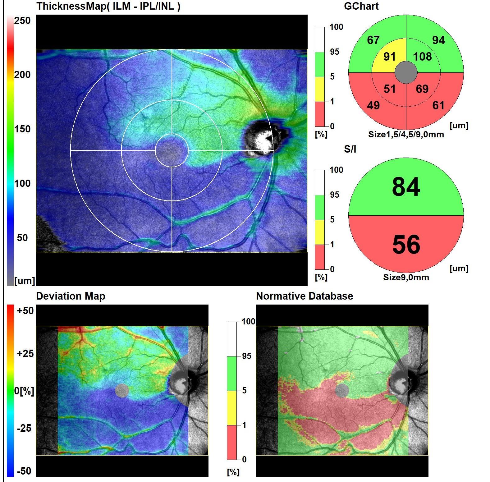
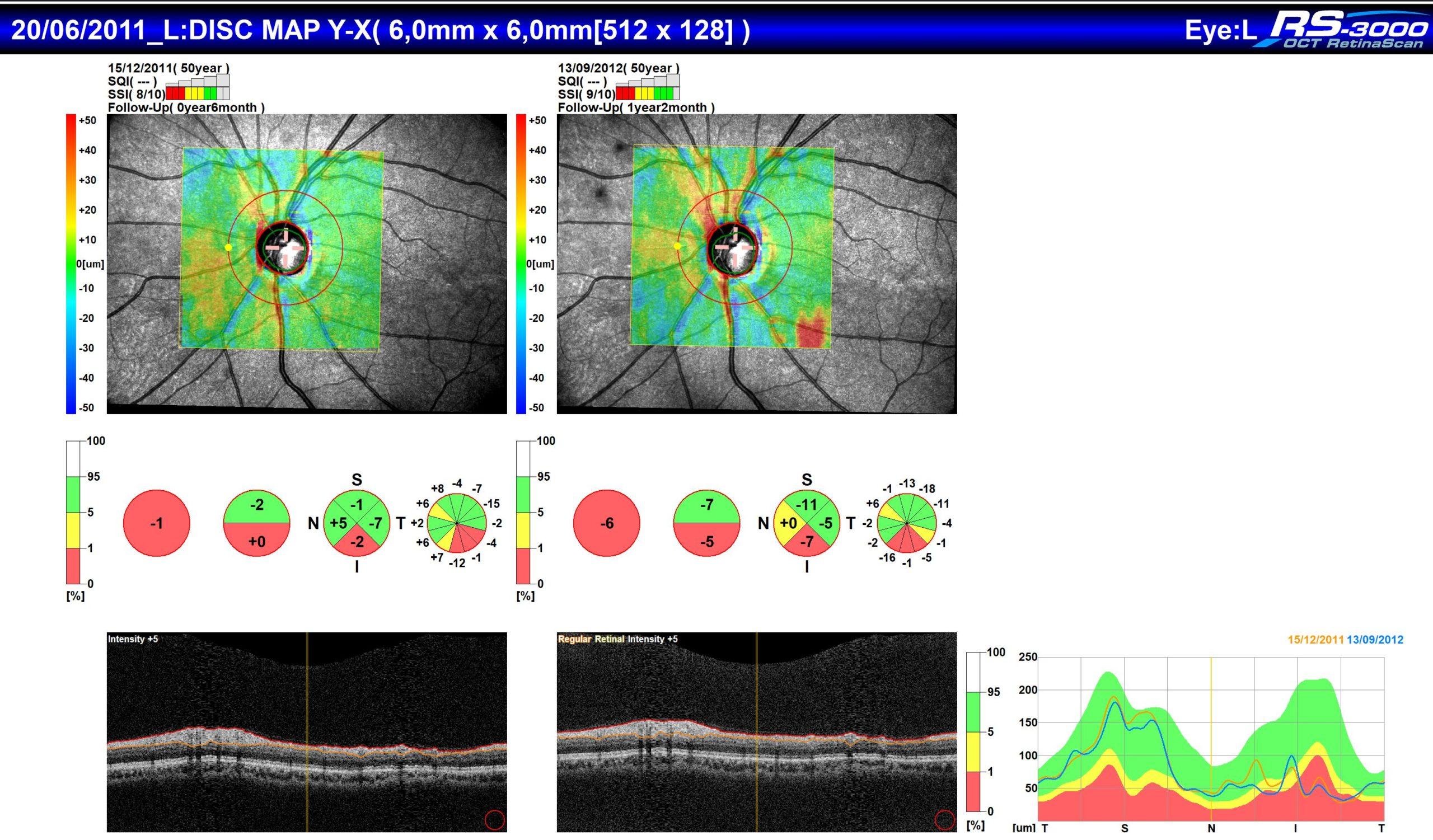
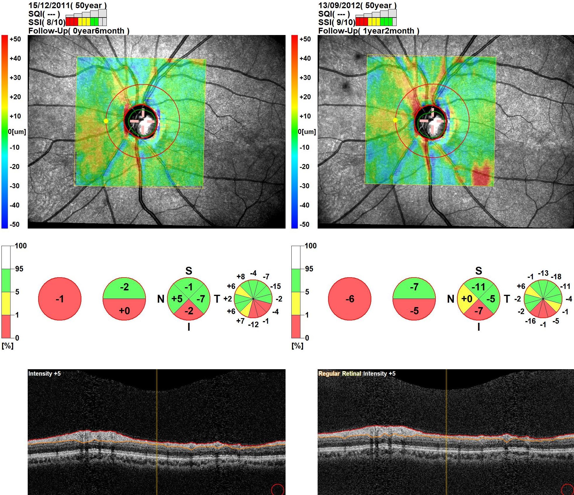
Thanks to this level of accuracy, the ophthalmologist can detect on these images even the finest pathological lesions of retina and choroid. By associating 2D sections, the OCT makes also possible rebuilding 3D volumes of the fundus, and this acquisition is essential to study retina thicknesses (oedemas and retinal detachments) and particularly the complex of ganglionic cells, in the glaucoma. Theses values are then compared in the patient following-up to monitor the evolution of the disease, to control the efficiency of the cure and to adjust the therapeutic strategy.
An additional lens moves the focal plane of the device to the anterior segment, thus making possible making an analysis of the section of the cornea and of the iridocorneal angle (pachymetry, cornea diseases and glaucomas by closing the angle).
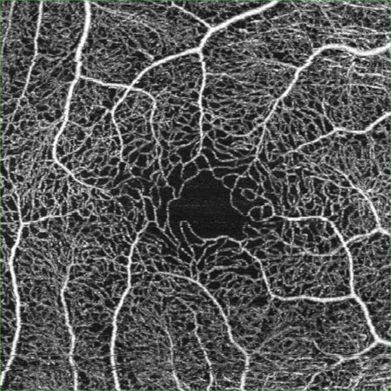
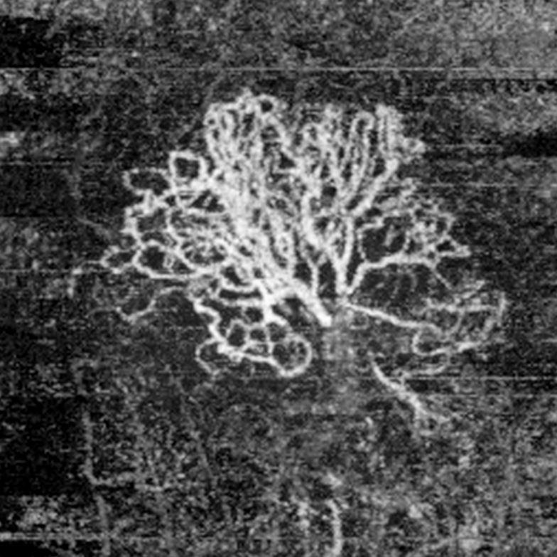
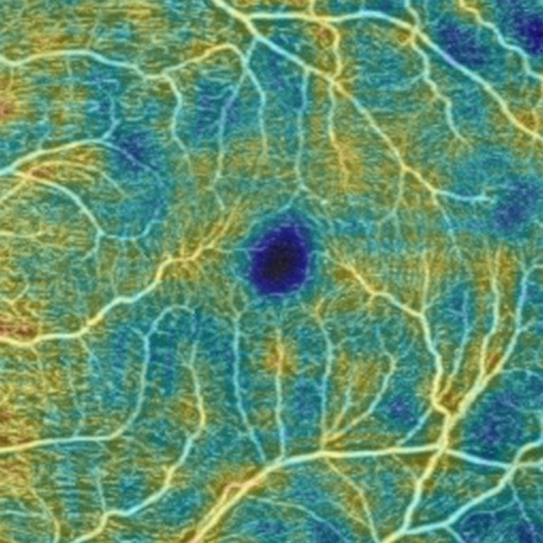
The evolution of the processing of the OCT signal has lead to a new analysis method of retinal-choroidal micro vascularisation, the OCT-Angiography.
Based upon a detection technique of the motion ('Motion contrast') and thanks to the acquisition of two OCT pictures on the same location but separated in time, the OCT-Angiography measures the motion of blood elements circulating in both retina and choroid.
Rebuilding a volume displays the grid of the vascular network in 3 dimensions. The user can then use this grid to analyse any section of any tissue thickness.
Unlike the injection angiography, this technique is not a dynamic one and its application field is still limited. Nevertheless, its main benefit is it is non-invasive (no risk of anaphylactic shock) and it can precisely locate the lesion(s) in tissue’s depth.
Therefore, the OCT-Angiography is regularly used but does not replace all the indications provided by the injection angiography.


Pictures courtesy: CARGO (CHU Strasbourg, France), CHIC (Creteil, France), Doctor Villalobos (Lima, Peru), Kittner Eye Center UNC (North Carolina, USA)
OCT B-Scan denoising is deep learning artificial intelligence software that enables high definition OCT B-Scan cutting from a single image. The processed images are thus of a quality equivalent to a high image summation although they are acquired in a minimum of time.
The NIDEK OCT B-Scan denoising software has been developed and trained on a large basis of OCT HD summed 120 times. For optimum efficiency, the software uses a deep learning process based on a Generative Adversarial Networks (GAN) system, a promising technology recognized as an effective approach in improving image clarity.
Whatever it is designed to analyse the posterior segment or the anterior segment, the OCT displays tissue’s layers with a high resolution, thus allowing a detailed structural analysis of imaged areas.
With a deep focus, the OCT of the posterior segment provides an image of the fundus, from vitreous to choroid. So all the lesions caused by retinal and choroidal pathologies can be detected to start detection, to make a diagnosis and perform the patient following-up.
There are different types of OCT of the posterior segment, depending on the wavelength used, on the related fundus visualisation system, on the acquisition technique of images and on related functionalities. Each type presents its own particular benefits.
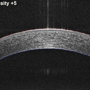
OCT’s of the anterior segment have an adaptive lens to move the focal plane to the level of the section analysis of the anterior part of the eye. For a better penetration, we have developed OCT’s dedicated to the anterior segment. They use a swept-source technology, better adapted to the imaging of the anterior chamber and make possible a detailed analysis of its structures.
Corneal pathologies, lenticular anomalies, implant positioning, opening of iridocorneal angle, etc. are then studied and tracked.
The OCT has revolutionised fundus analysis as fundus was previously impossible to reach in vivo and makes possible making a very precise diagnosis and following-up of retinal-choroidal pathologies. It has become an essential routine examination.

Examen IR facile à réaliser avec une grande quantité d’information obtenue en peu de temps.

2D section results or 3D reconstruction with analysis of thicknesses, including the complex of ganglionic cells.

Using high-contrast SLO images or colour photo, complete the section analysis with a front analysis.

Imaging of retina and choroid vascular network to detect anomalies and infusion changes.
You have a project? You want a quotation? You have questions about our products? Feel free to ask your technical sales representative.
Reliability and
safety
NIDEK develops its top-of-the-range products to improve visual health through an approach based on strict criteria: safety, reliability, durability, continuous quality controls and certifications.
Technologies and
innovations
NIDEK meets technical challenges by keeping constantly informed of the innovations of eye imaging systems, using the expertise of professionals and the progresses of research.
Services and
guarantees
NIDEK commits itself to providing services to its customers, from the installation of an activity to the authorised training of teams, and to offering long-time measurable guarantees.
Sucy-en-Brie, le 30 avril 2026 – NIDEK France, leader mondial en équipements d’optique et d’ophtalmologie,
Du 9 au 11 mai 2026, NIDEK vous donne rendez-vous au Palais des Congrès de
NIDEK redéfinît les standards de meulage avec la nouvelle série LEXCE Plus, des meuleuses multifonctions ultra-compactes
1 : Notre bureau d’études pour vous aider sur vos implantations 2 : Espace préconsultation
Du 9 au 11 mai 2026, NIDEK vous donne rendez-vous au Palais des Congrès de
NIDEK SA Lauréate du Mois de l’Économie de la Ville de Saint-Priest Nous sommes heureux
Le Silmo Paris 2025, rendez-vous incontournable de l’optique et de la lunetterie, se tiendra du
Saint-Priest (69), le 12 juin 2025 – NIDEK SA, filiale française du groupe japonais NIDEK,
Ce site est réservé aux professionnels de santé