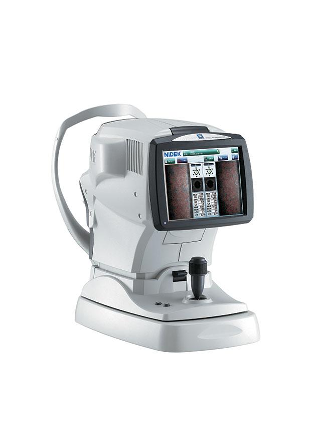
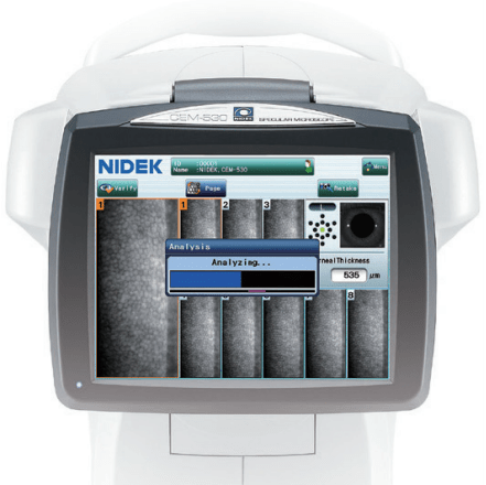
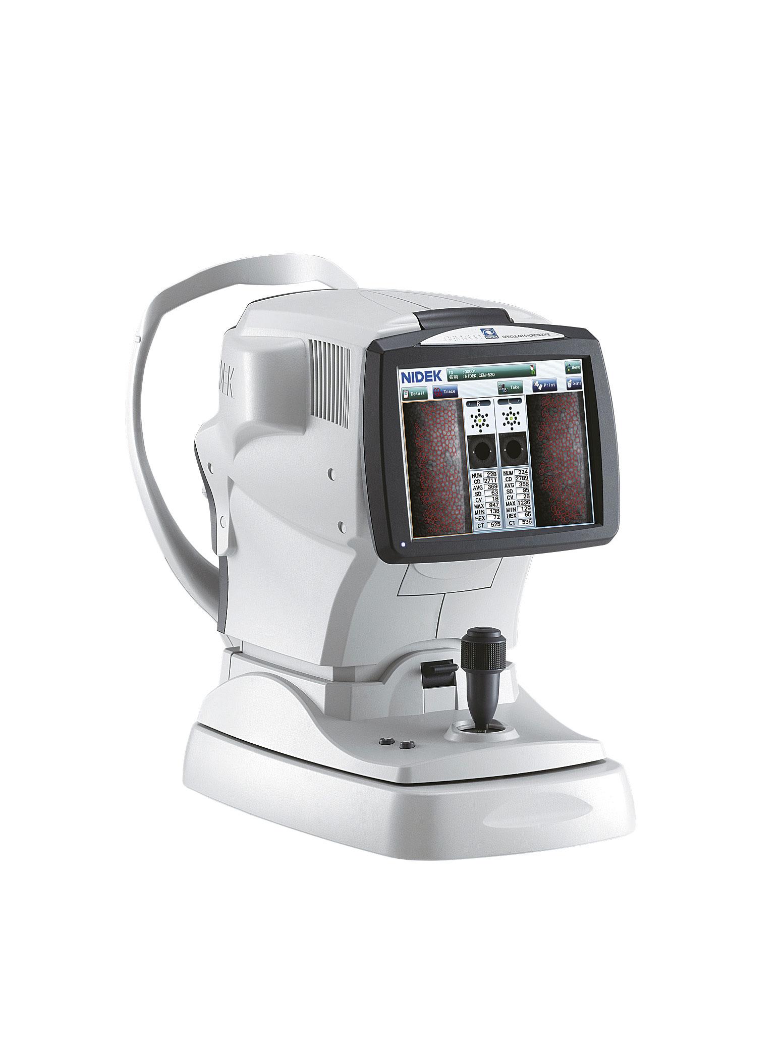
Simple and intuitive navigation from its large touch screen
Quick acquisition of the measurements
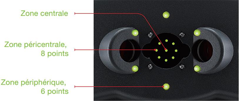
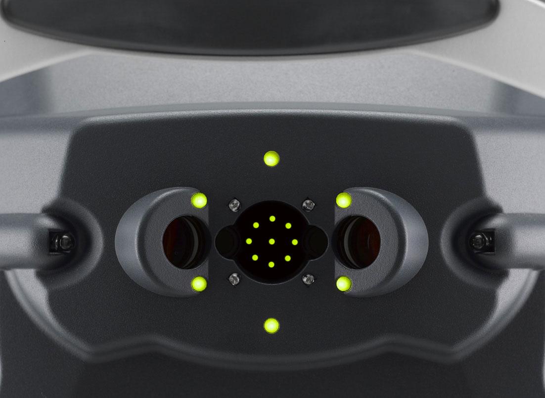
Measurement of the central area, of pericentral areas (8 points) and peripheral areas (6 points)
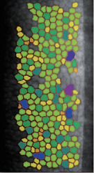
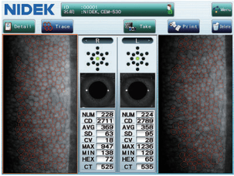
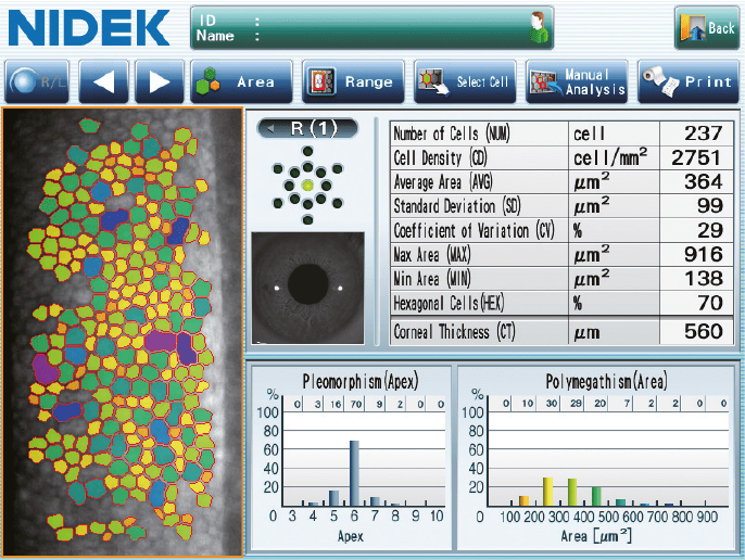
Built-in analysis software with two-side displays, automatic and manual counting
Transmission of the results to the patient management softwares
Viewer CEM-530 is available (optional) through the NAVIS-EX imaging software (patient data management software)
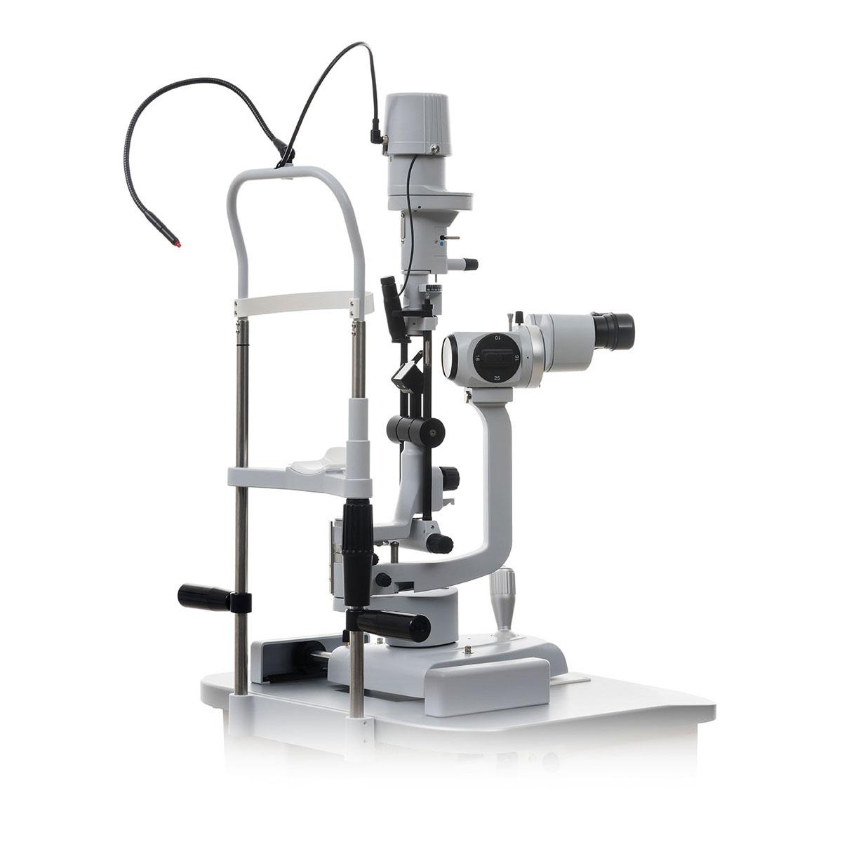
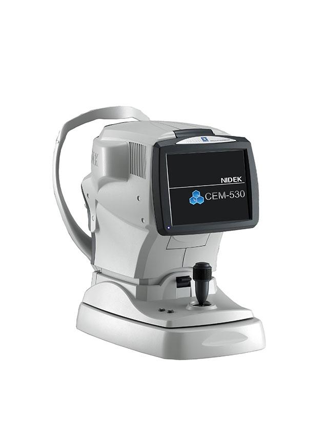
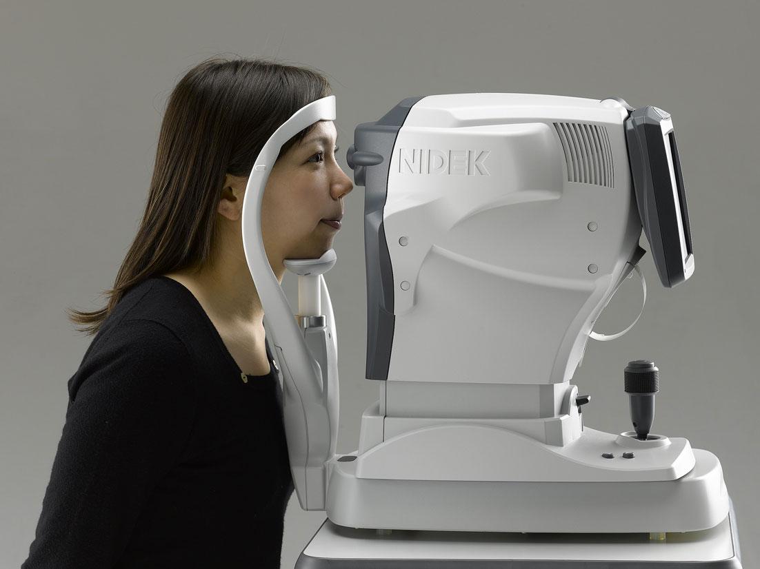
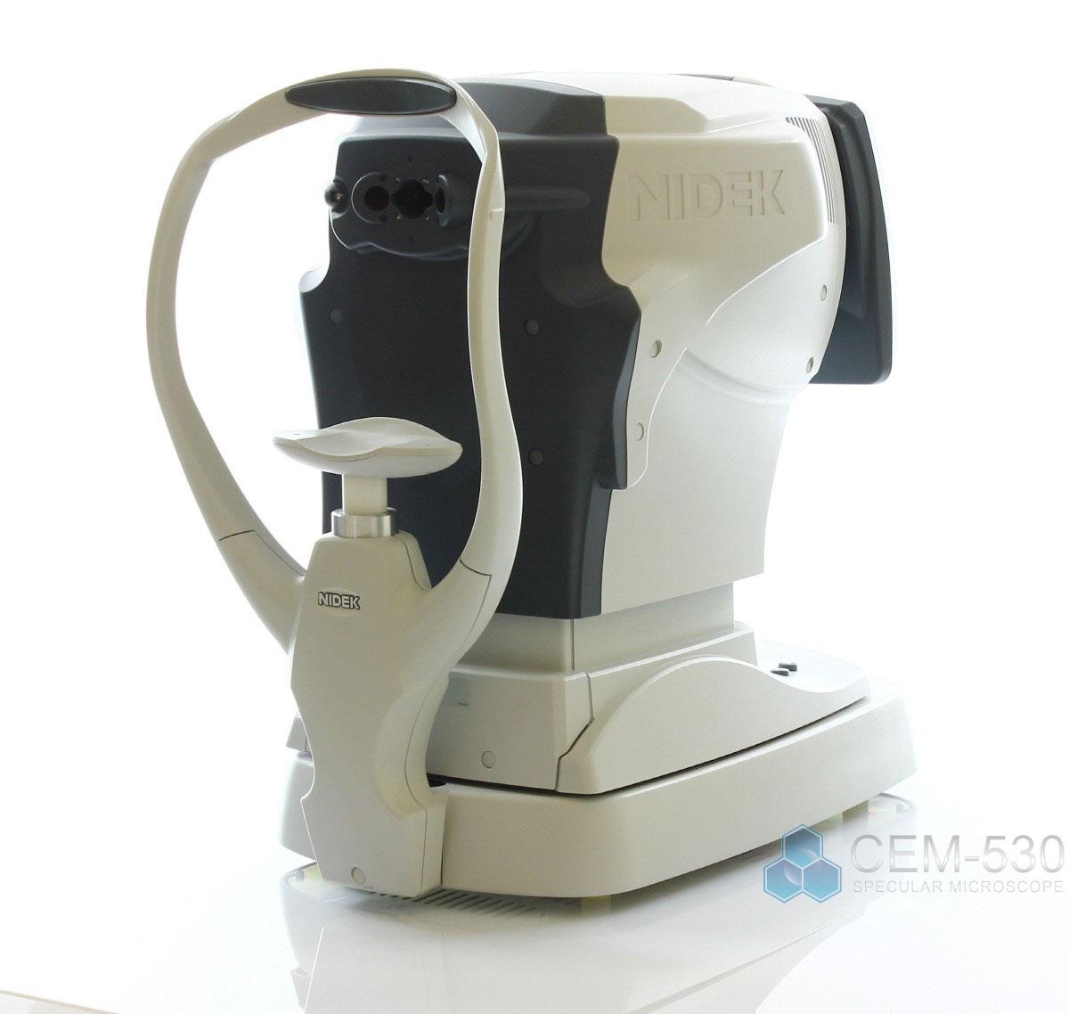
Specular microscopy
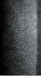
The specular microscope is a non-invasive imaging and analysis device of the corneal endothelial layer. The specular microscopy is an examination of endothelial cells using the specular reflexion resulting from the interface between endothelial cells and aqueous humour.
The different analysis zones
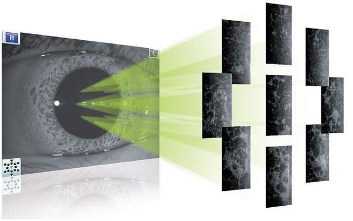
The specular microscope CEM-530 performs central, paracentral (diameter: 1.3 mm) and peripheral (diameter: 7.3 mm) conventional specular microscopies. The combination of central, paracentral and peripheral imaging provides an overview that can be used to make a detailed morphological and quantitative evaluation of the endothelial layer.
Paracentral images are captured from 8 points under a visual angle of 5° on a diameter of 1.3 mm to perform an improved evaluation surrounding the central appearance of the cornea.
Peripheral images are captured from 6 points under a visual angle of 27° on a diameter of 7.3 mm to perform an improved evaluation of the peripheral zone of the cornea.
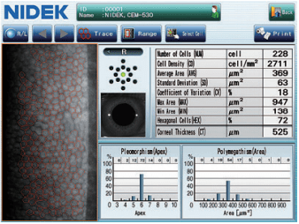
Quantitative & qualitative analysis
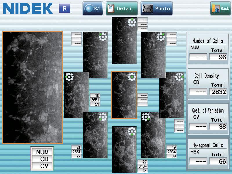
A series of 16 images are captured. These images are automatically sorted out depending on their quality. The built-in software identifies the image used to optimise the counting and perform the analysis of endothelial cells. Using the data is simple and quick.
Analysing the endothelium specular image consists in evaluating the appearance of cells covering this specific cornea layer to bring out anomalies such as drops or keratic precipitates. Very often, the operator has to count the number of cells in a defined area while analysing the distribution of the cell sizes.
Suitable for all practices
To perform a detailed analysis, the user can mark out the observation zone and take out of the counting the parts of the images that are inappropriate.
Moreover, a measurement of the central corneal thickness completes the data captured by the microscope.
In order to adapt to types of working organisation, connecting the CEM-530 to a network is possible. So it makes possible directly triggering from the machine the printing to a printer connected to the computer or directly sending the digital reports to a patient management software.
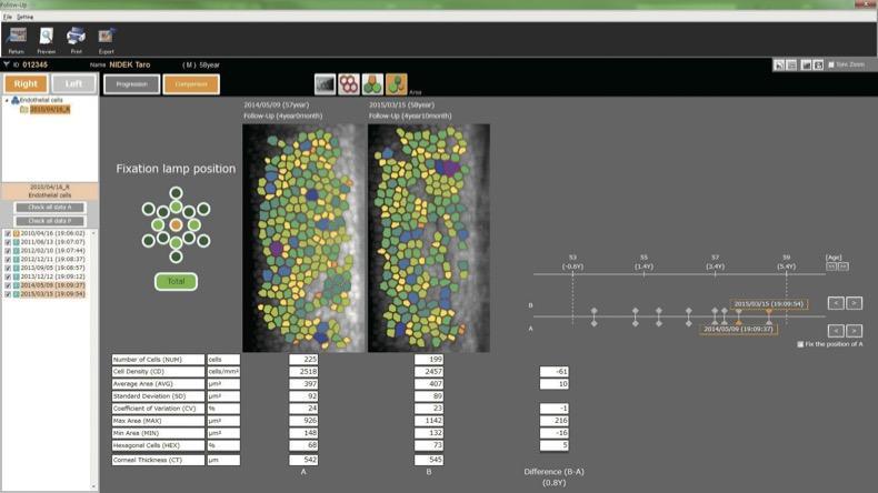
Viewer CEM-530 is available (optional) through the NAVIS-EX imaging software (patient data management software). The viewer CEM-530 allows a following-up of the counting of endothelium cells over time
NIDEK develops its top-of-the-range products to improve visual health through an approach based on strict criteria: safety, reliability, durability, continuous quality controls and certifications.
Technologies and innovations
NIDEK meets technical challenges by keeping constantly informed of the innovations of eye imaging systems, using the expertise of professionals and the progresses of research.
Services and guarantees
NIDEK commits itself to providing services to its customers, from the installation of an activity to the authorised training of teams, and to offering long-time measurable guarantees.
Nous utilisons des cookies pour optimiser notre site web et notre service.
Fonctionnel
Always active
Le stockage ou l’accès technique est strictement nécessaire dans la finalité d’intérêt légitime de permettre l’utilisation d’un service spécifique explicitement demandé par l’abonné ou l’utilisateur, ou dans le seul but d’effectuer la transmission d’une communication sur un réseau de communications électroniques.
Préférences
Le stockage ou l’accès technique est nécessaire dans la finalité d’intérêt légitime de stocker des préférences qui ne sont pas demandées par l’abonné ou l’utilisateur.
Statistiques
Le stockage ou l’accès technique qui est utilisé exclusivement à des fins statistiques.Le stockage ou l’accès technique qui est utilisé exclusivement dans des finalités statistiques anonymes. En l’absence d’une assignation à comparaître, d’une conformité volontaire de la part de votre fournisseur d’accès à internet ou d’enregistrements supplémentaires provenant d’une tierce partie, les informations stockées ou extraites à cette seule fin ne peuvent généralement pas être utilisées pour vous identifier.
Marketing
Le stockage ou l’accès technique est nécessaire pour créer des profils d’utilisateurs afin d’envoyer des publicités, ou pour suivre l’utilisateur sur un site web ou sur plusieurs sites web ayant des finalités marketing similaires.